Hispanopedia Wiki

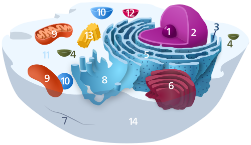
Mitochondrial DNA (mDNA or mtDNA) is the DNA located in the mitochondriaorganelles in a eukaryotic cell that converts chemical energy from organic compounds into adenosine triphosphate (ATP). Mitochondrial DNA is a small portion of the DNA contained in a eukaryotic cell; most of the DNA is in the cell nucleus, and, in plants and algae, the DNA also is found in plastids, such as chloroplasts.[3] Mitochondrial DNA is responsible for coding of 13 essential subunits of the complex oxidative phosphorylation (OXPHOS) system which has a role in cellular energy conversion.[4]
Human mitochondrial DNA was the first significant part of the human genome to be sequenced.[5] This sequencing revealed that human mtDNA has 16,569 base pairs and encodes 13 proteins. As in other vertebrates, the human mitochondrial genetic code differs slightly from nuclear DNA.[6]
Since animal mtDNA evolves faster than nuclear genetic markers,[7][8][9] it represents a mainstay of phylogenetics and evolutionary biology. It also permits tracing the relationships of populations, and so has become important in anthropology and biogeography.
Origin
Se cree que el ADN nuclear y el mitocondrial tienen orígenes evolutivos separados , y que el ADNmt deriva de los genomas circulares de bacterias que fueron engullidas por los ancestros de las células eucariotas modernas. Esta teoría se denomina teoría endosimbiótica . En las células de los organismos actuales, la gran mayoría de las proteínas mitocondriales (aproximadamente 1500 tipos diferentes en mamíferos ) están codificadas por el ADN nuclear , pero se cree que los genes de algunas, si no de la mayoría, de ellas son de origen bacteriano, habiéndose transferido al núcleo eucariota durante la evolución . [ 10 ]
Se debaten las razones por las que las mitocondrias han conservado algunos genes. La existencia en algunas especies de orgánulos derivados de mitocondrias que carecen de genoma [ 11 ] sugiere que es posible la pérdida completa de genes, y la transferencia de genes mitocondriales al núcleo tiene varias ventajas. [ 12 ] La dificultad de dirigir productos proteicos hidrofóbicos producidos remotamente a la mitocondria es una hipótesis para explicar por qué se conservan algunos genes en el ADNmt; [ 13 ] la colocalización para la regulación redox es otra, citando la conveniencia del control localizado sobre la maquinaria mitocondrial. [ 14 ] Un análisis reciente de una amplia gama de genomas de ADNmt sugiere que ambas características pueden determinar la retención de genes mitocondriales. [ 10 ]
Estructura y diversidad del genoma
En todos los organismos, existen seis tipos principales de genoma mitocondrial, clasificados por su estructura (es decir, circular versus lineal), tamaño, presencia de intrones o estructuras similares a plásmidos , y si el material genético es una molécula singular o una colección de moléculas homogéneas o heterogéneas . [ 15 ]
En muchos organismos unicelulares (por ejemplo, el ciliado Tetrahymena y el alga verde Chlamydomonas reinhardtii ), y en casos raros también en organismos multicelulares (por ejemplo, en algunas especies de Cnidaria ), el ADNmt es ADN lineal . La mayoría de estos ADNmt lineales poseen telómeros independientes de la telomerasa (es decir, los extremos del ADN lineal) con diferentes modos de replicación, lo que los ha convertido en objetos de investigación interesantes porque muchos de estos organismos unicelulares con ADNmt lineal son patógenos conocidos . [ 16 ]
Animales
Most (bilaterian) animals have a circular mitochondrial genome. Medusozoa and calcareaclades however include species with linear mitochondrial chromosomes.[17] With a few exceptions, animals have 37 genes in their mitochondrial DNA: 13 for proteins, 22 for tRNAs, and 2 for rRNAs.[18]
Mitochondrial genomes for animals average about 16,000 base pairs in length.[18] The anemone Isarachnanthus nocturnus has the largest mitochondrial genome of any animal at 80,923 bp.[19] The smallest known mitochondrial genome in animals belongs to the comb jelly Vallicula multiformis, which consist of 9,961 bp.[20]
In February 2020, a jellyfish-related parasite – Henneguya salminicola – was discovered that lacks a mitochondrial genome but retains structures deemed mitochondrion-related organelles. Moreover, nuclear DNA genes involved in aerobic respiration and mitochondrial DNA replication and transcription were either absent or present only as pseudogenes. This is the first multicellular organism known to have this absence of aerobic respiration and live completely free of oxygen dependency.[21][22]
The Armadillidium vulgare has a uniquely large genome when compared to other crustacean average about 30 kB greater allowing it to be a model organism in mtDNA research[23].
Plants and fungi
There are three different mitochondrial genome types in plants and fungi. The first type is a circular genome that has introns (type 2) and may range from 19 to 1000 kbp in length. The second genome type is a circular genome (about 20–1000 kbp) that also has a plasmid-like structure (1 kb) (type 3). The final genome type found in plants and fungi is a linear genome made up of homogeneous DNA molecules (type 5).[24][25][26]
Great variation in mtDNA gene content and size exists among fungi and plants, although there appears to be a core subset of genes present in all eukaryotes (except for the few that have no mitochondria at all).[10] In Fungi, however, there is no single gene shared among all mitogenomes.[27] Some plant species have enormous mitochondrial genomes, with Silene conica mtDNA containing as many as 11,300,000 base pairs.[28] However, even those huge mtDNAs contain the same number and kinds of genes as related plants with much smaller mtDNAs.[29] The genome of the mitochondrion of the cucumber (Cucumis sativus) consists of three circular chromosomes (lengths 1556, 84 and 45 kilobases), which are entirely or largely autonomous with regard to their replication.[30]
Protists
Protists contain the most diverse mitochondrial genomes, with five different types found in this kingdom. Type 2, type 3, and type 5 of the plant and fungal genomes also exist in some protists, as do two unique genome types. One of these unique types is a heterogeneous collection of circular DNA molecules (type 4) while the other is a heterogeneous collection of linear molecules (type 6). Genome types 4 and 6 each range from 1–200 kbp in size.
The smallest mitochondrial genome sequenced to date is the 5,967 bp mtDNA of the parasite Plasmodium falciparum.[31][32]
Endosymbiotic gene transfer, the process by which genes that were coded in the mitochondrial genome are transferred to the cell's main genome, likely explains why more complex organisms such as humans have smaller mitochondrial genomes than simpler organisms such as protists.
Replication
The two strands of the human mitochondrial DNA are distinguished as the heavy strand and the light strand.[33] The regulation of mitochondrial DNA replication and transcription initiation is located in a single intergenic noncoding region (NCR).[33] In humans, the 1,100 base pair NCR region contains three promoters of two L-strand promoters (LSP and LSP2) and one H-strand promoter (HSP).[34] Unlike bidirectional and specific origin initiation of nuclear DNA replication, mitochondrial DNA has two strand-specific, unidirectional origins of replication of the leading H strand (OH) which located in NCR and the lagging L strand (OL) which located in the tRNA gene cluster.[35]
Mitochondrial DNA is replicated by the DNA polymerase gamma complex which is composed of a 140 kDa catalytic DNA polymerase encoded by the POLG gene and two 55 kDa accessory subunits encoded by the POLG2 gene.[36] The replisome machinery is formed by DNA polymerase, TWINKLE and mitochondrial SSB proteins. TWINKLE is a helicase, which unwinds short stretches of dsDNA in the 5' to 3' direction.[37] Mitochondrial single-stranded DNA binding protein (SSB) coordinates its activity with the POLG during DNA replication.[38] All these polypeptides are encoded in the nuclear genome.
During embryogenesis, replication of mtDNA is strictly down-regulated from the fertilized oocyte through the preimplantation embryo.[39] The resulting reduction in per-cell copy number of mtDNA plays a role in the mitochondrial bottleneck, exploiting cell-to-cell variability to ameliorate the inheritance of damaging mutations.[40] According to Justin St. John and colleagues, "At the blastocyst stage, the onset of mtDNA replication is specific to the cells of the trophectoderm.[39] In contrast, the cells of the inner cell mass restrict mtDNA replication until they receive the signals to differentiate to specific cell types."[39]
DNA repair
Although several DNA repair pathways have been reported to occur in the mitochondria, currently the base excision repair pathway is the pathway most comprehensively described.[41] Proteins that are employed in the maintenance of mitochondrial DNA are encoded by nuclear genes and translocated to the mitochondria.[41] The mitochondria of human cells are capable of repairing DNAbase pair mismatches by a pathway that is distinct from the DNA mismatch repair pathway of the nucleus.[42] This distinct mitochondrial pathway includes the activity of the Y box binding protein 1 (designated YB-1 or YBX1), that likely acts in the mismatch binding and recognition steps of mismatch repair.[42] DNA repair mechanisms specific to the mitochondria may reflect the proximity of the mitochondrial DNA to the oxidative phosphorylation system and consequently to the DNA-damaging reactive oxygen species formed during ATP production.[43]

Genes on the human mtDNA and their transcription

The two strands of the human mitochondrial DNA are distinguished as the heavy strand and the light strand. The heavy strand is rich in guanine and encodes 12 subunits of the oxidative phosphorylation system, two ribosomal RNAs (12S and 16S), and 14 transfer RNAs (tRNAs). The light strand encodes one subunit and 8 tRNAs. So, altogether mtDNA encodes for two rRNAs, 22 tRNAs, and 13 protein subunits, all of which are involved in the oxidative phosphorylation process.[46][47]
- The complete sequence of the human mitochondrial DNA in graphic form
Between most (but not all) protein-coding regions, tRNAs are present (see the human mitochondrial genome map). During transcription, the tRNAs acquire their characteristic L-shape that gets recognized and cleaved by specific enzymes. With the mitochondrial RNA processing, individual mRNA, rRNA, and tRNA sequences are released from the primary transcript.[49] Folded tRNAs therefore act as secondary structure punctuations.[50]
Transcription is done by the single-subunit mitochondrial RNA polymerase (POLRMT). In association with two of accessory factors, mitochondrial transcription factor A (TFAM) and mitochondrial transcription factor B2 (TFB2M), the POLRMT complex recognizes promoters and initiates transcription.[51] Transcription resulted in polycistronic transcripts that are processed in discrete mitochondrial RNA granules into individual mRNAs, tRNAs, and rRNAs.[52]
Regulation of transcription
The promoters for the initiation of the transcription of the heavy and light strands are located in the main non-coding region of the mtDNA called the displacement loop, the D-loop.[46] There is evidence that the transcription of the mitochondrial rRNAs is regulated by the heavy-strand promoter 1 (HSP1), and the transcription of the polycistronic transcripts coding for the protein subunits are regulated by HSP2.[46]
Measurement of the levels of the mtDNA-encoded RNAs in bovine tissues has shown that there are major differences in the expression of the mitochondrial RNAs relative to total tissue RNA.[53] Among the 12 tissues examined the highest level of expression was observed in the heart, followed by brain and steroidogenic tissue samples.[53]
As demonstrated by the effect of the trophic hormone ACTH on adrenal cortex cells, the expression of the mitochondrial genes may be strongly regulated by external factors, apparently to enhance the synthesis of mitochondrial proteins necessary for energy production.[53] Interestingly, while the expression of protein-encoding genes was stimulated by ACTH, the levels of the mitochondrial 16S rRNA showed no significant change.[53]
Mitochondrial inheritance
In most multicellular organisms, mtDNA is inherited from the mother (maternally inherited). Mechanisms for this include simple dilution (an egg contains on average 200,000 mtDNA molecules, whereas a healthy human sperm has been reported to contain on average 5 molecules),[54][55] degradation of sperm mtDNA in the male genital tract and the fertilized egg; and, at least in a few organisms, failure of sperm mtDNA to enter the egg. Whatever the mechanism, this single parent (uniparental inheritance) pattern of mtDNA inheritance is found in most animals, most plants, and also in fungi.[56]
In a study published in 2018, human babies were reported to inherit mtDNA from both their fathers and their mothers resulting in mtDNA heteroplasmy,[57] a finding that has been rejected by other scientists.[58][59][60]
Female inheritance
In sexual reproduction, mitochondria are normally inherited exclusively from the mother; the mitochondria in mammalian sperm are usually destroyed by the egg cell after fertilization. Also, mitochondria are present solely in the midpiece, which is used for propelling the sperm cells, and sometimes the midpiece, along with the tail, is lost during fertilization. In 1999 it was reported that paternal sperm mitochondria (containing mtDNA) are marked with ubiquitin to select them for later destruction inside the embryo.[61] Some in vitro fertilization techniques, particularly injecting a sperm into an oocyte, may interfere with this.
The fact that mitochondrial DNA is mostly maternally inherited enables genealogical researchers to trace maternal lineage far back in time. ( Y-chromosomal DNA , paternally inherited, is used in an analogous way to determine the patrilineal history.) This is usually accomplished on human mitochondrial DNA by sequencing the hypervariable control regions (HVR1 or HVR2), and sometimes the complete molecule of the mitochondrial DNA, as a genealogical DNA test . [ 62 ] HVR1, for example, consists of about 440 base pairs. These 440 base pairs are compared to the same regions of other individuals (either specific people or subjects in a database) to determine maternal lineage. Most often, the comparison is made with the revised Cambridge Reference Sequence . Vilà et al. have published studies tracing the matrilineal descent of domestic dogs from wolves. [ 63 ] El concepto de la Eva mitocondrial se basa en el mismo tipo de análisis, tratando de descubrir el origen de la humanidad rastreando el linaje hacia atrás en el tiempo.
El cuello de botella mitocondrial
Se espera que las entidades sujetas a herencia uniparental y con poca o ninguna recombinación estén sujetas al trinquete de Muller , la acumulación de mutaciones deletéreas hasta la pérdida de funcionalidad. Las poblaciones animales de mitocondrias evitan esto mediante un proceso de desarrollo conocido como cuello de botella del ADNmt . El cuello de botella aprovecha los procesos aleatorios en la célula para aumentar la variabilidad célula a célula en la carga mutante a medida que se desarrolla un organismo: un solo óvulo con cierta proporción de ADNmt mutante produce un embrión en el que diferentes células tienen diferentes cargas mutantes. La selección a nivel celular puede entonces actuar para eliminar aquellas células con más ADNmt mutante, lo que lleva a una estabilización o reducción de la carga mutante entre generaciones. El mecanismo subyacente al cuello de botella es objeto de debate, [ 64 ] [ 65 ] [ 66 ] [ 67 ] y un metaestudio matemático y experimental reciente proporciona evidencia de una combinación de la partición aleatoria de ADNmt en las divisiones celulares y el recambio aleatorio de moléculas de ADNmt dentro de la célula. [ 40 ]
Herencia masculina
Male mitochondrial DNA inheritance has been discovered in Plymouth Rock chickens.[68] Evidence supports rare instances of male mitochondrial inheritance in some mammals as well. Specifically, documented occurrences exist for mice,[69][70] where the male-inherited mitochondria were subsequently rejected. It has also been found in sheep,[71] and in cloned cattle.[72] Rare cases of male mitochondrial inheritance have been documented in humans.[73][74][75][57] Although many of these cases involve cloned embryos or subsequent rejection of the paternal mitochondria, others document in vivo inheritance and persistence under lab conditions.
Doubly uniparental inheritance of mtDNA is observed in bivalve mollusks. In those species, females have only one type of mtDNA (F), whereas males have F-type mtDNA in their somatic cells, but M-type mtDNA (which can be as much as 30% divergent) in germline cells.[76] Paternally inherited mitochondria have additionally been reported in some insects such as fruit flies,[77][78]honeybees,[79] and periodical cicadas.[80]
Mitochondrial donation
An IVF technique known as mitochondrial donation or mitochondrial replacement therapy (MRT) results in offspring containing mtDNA from a donor female, and nuclear DNA from the mother and father. In the spindle transfer procedure, the nucleus of an egg is inserted into the cytoplasm of an egg from a donor female which has had its nucleus removed but still contains the donor female's mtDNA. The composite egg is then fertilized with the male's sperm. The procedure is used when a woman with genetically defective mitochondria wishes to procreate and produce offspring with healthy mitochondria.[81] The first known child to be born as a result of mitochondrial donation was a boy born to a Jordanian couple in Mexico on 6 April 2016.[82]
Mutations and disease


Susceptibility
The concept that mtDNA is particularly susceptible to reactive oxygen species generated by the respiratory chain due to its proximity remains controversial.[83] mtDNA does not accumulate any more oxidative base damage than nuclear DNA.[84] It has been reported that at least some types of oxidative DNA damage are repaired more efficiently in mitochondria than they are in the nucleus.[85] mtDNA is packaged with proteins which appear to be as protective as proteins of the nuclear chromatin.[86] Moreover, mitochondria evolved a unique mechanism which maintains mtDNA integrity through degradation of excessively damaged genomes followed by replication of intact/repaired mtDNA. This mechanism is not present in the nucleus and is enabled by multiple copies of mtDNA present in mitochondria.[87] The outcome of mutation in mtDNA may be an alteration in the coding instructions for some proteins,[88] which may have an effect on organism metabolism and/or fitness.
Genetic illness
Mutations of mitochondrial DNA can lead to a number of illnesses including exercise intolerance and Kearns–Sayre syndrome (KSS), which causes a person to lose full function of heart, eye, and muscle movements. Some evidence suggests that they might be major contributors to the aging process and age-associated pathologies.[89] Particularly in the context of disease, the proportion of mutant mtDNA molecules in a cell is termed heteroplasmy. The within-cell and between-cell distributions of heteroplasmy dictate the onset and severity of disease[90] and are influenced by complicated stochastic processes within the cell and during development.[40][91]
Mutations in mitochondrial tRNAs can be responsible for severe diseases like the MELAS and MERRF syndromes.[92]
Mutations in nuclear genes that encode proteins that mitochondria use can also contribute to mitochondrial diseases. These diseases do not follow mitochondrial inheritance patterns but instead follow Mendelian inheritance patterns.[93]
Use in disease diagnosis
Recently a mutation in mtDNA has been used to help diagnose prostate cancer in patients with negative prostate biopsy.[94][95] mtDNA alterations can be detected in the bio-fluids of patients with cancer.[96] mtDNA is characterized by the high rate of polymorphisms and mutations. Some of these are increasingly recognized as an important cause of human pathology such as oxidative phosphorylation (OXPHOS) disorders, maternally inherited diabetes and deafness (MIDD), Type 2 diabetes mellitus, Neurodegenerative disease, heart failure, and cancer.
Relationship with ageing
Though the idea is controversial, some evidence suggests a link between aging and mitochondrial genome dysfunction. [ 97 ] In essence, mutations in mtDNA upset a careful balance of reactive oxygen species (ROS) production and enzymatic ROS scavenging (by enzymes like superoxide dismutase , catalase , glutathione peroxidase and others). However, some mutations that increase ROS production (eg, by reducing antioxidant defenses) in worms increase, rather than decrease, their longevity. [ 83 ] Also, naked mole rats , rodents about the size of mice , live about eight times longer than mice despite having reduced, compared to mice, antioxidant defenses and increased oxidative damage to biomolecules. [ 98 ] Once, there was thought to be a positive feedback loop at work (a 'Vicious Cycle'); as mitochondrial DNA accumulates genetic damage caused by free radicals, the mitochondria lose function and leak free radicals into the cytosol . A decrease in mitochondrial function reduces overall metabolic efficiency. [ 99 ] However, this concept was conclusively disproved when it was demonstrated that mice, which were genetically altered to accumulate mtDNA mutations at an accelerated rate to age prematurely, but their tissues do not produce more ROS as predicted by the 'Vicious Cycle' hypothesis. [ 100 ] Supporting a link between longevity and mitochondrial DNA, some studies have found correlations between biochemical properties of the mitochondrial DNA and the longevity of species. [ 101 ] The application of a mitochondrial-specific ROS scavenger, which lead to a significant longevity of the mice studied, [ 102 ] suggests that mitochondria may still be well-implicated in ageing. Extensive research is being conducted to further investigate this link and methods to combat ageing. Presently, gene therapy and nutraceutical supplementation are popular areas of ongoing research. [ 103 ] [ 104 ] Bjelakovic et al. analizaron los resultados de 78 estudios entre 1977 y 2012, que involucraron a un total de 296.707 participantes, y concluyeron que los suplementos antioxidantes no reducen la mortalidad por todas las causas ni prolongan la esperanza de vida, mientras que algunos de ellos, como el betacaroteno, la vitamina E y las dosis más altas de vitamina A, pueden incluso aumentar la mortalidad. [ 105 ] In a recent study, it was shown that dietary restriction can reverse ageing alterations by affecting the accumulation of mtDNA damage in several organs of rats. For example, dietary restriction prevented age-related accumulation of mtDNA damage in the cortex and decreased it in the lung and testis.[106]
Neurodegenerative diseases
Increased mtDNA damage is a feature of several neurodegenerative diseases.
The brains of individuals with Alzheimer's disease have elevated levels of oxidative DNA damage in both nuclear DNA and mtDNA, but the mtDNA has approximately 10-fold higher levels than nuclear DNA.[107] It has been proposed that aged mitochondria is the critical factor in the origin of neurodegeneration in Alzheimer's disease.[108] Analysis of the brains of AD patients suggested an impaired function of the DNA repair pathway, which would cause reduce the overall quality of mtDNA.[109]
In Huntington's disease, mutant huntingtin protein causes mitochondrial dysfunction involving inhibition of mitochondrialelectron transport, higher levels of reactive oxygen species and increased oxidative stress.[110] Mutant huntingtin protein promotes oxidative damage to mtDNA, as well as nuclear DNA, that may contribute to Huntington's disease pathology.[111]
The DNA oxidation product 8-oxoguanine (8-oxoG) is a well-established marker of oxidative DNA damage. In persons with amyotrophic lateral sclerosis (ALS), the enzymes that normally repair 8-oxoG DNA damages in the mtDNA of spinal motor neurons are impaired.[112] Thus oxidative damage to mtDNA of motor neurons may be a significant factor in the etiology of ALS.
Correlation of the mtDNA base composition with animal life spans

Durante la última década, un grupo de investigación israelí liderado por el profesor Vadim Fraifeld ha demostrado que existen correlaciones fuertes y significativas entre la composición de bases del ADNmt y la longevidad máxima específica de cada especie animal. [ 113 ] [ 114 ] [ 115 ] Como se demuestra en su trabajo, un mayor contenido de guanina + citosina del ADNmt ( GC% ) se asocia fuertemente con una mayor longevidad máxima en todas las especies animales. Una observación adicional es que la correlación del GC% del ADNmt con la longevidad máxima es independiente de la conocida correlación entre la tasa metabólica de las especies animales y la longevidad máxima. El GC% del ADNmt y la tasa metabólica basal explican las diferencias en la longevidad máxima de las especies animales de manera multiplicativa (es decir, longevidad máxima de la especie = su GC% del ADNmt * tasa metabólica). [ 114 ] Para apoyar a la comunidad científica en la realización de análisis comparativos entre las características del ADNmt y la longevidad en los animales, se creó una base de datos dedicada llamada MitoAge . [ 116 ]
El espectro mutacional del ADN mitocondrial es sensible a los rasgos de la historia de vida específicos de cada especie.
De novo mutations arise either due to mistakes during DNA replication or due to unrepaired damage caused in turn by endogenous and exogenous mutagens. It has been long believed that mtDNA can be particularly sensitive to damage caused by reactive oxygen species (ROS), however, G>T substitutions, the hallmark of the oxidative damage in the nuclear genome, are very rare in mtDNA and do not increase with age. Comparing the mtDNA mutational spectra of hundreds of mammalian species, it has been recently demonstrated that species with extended lifespans have an increased rate of A>G substitutions on single-stranded heavy chains. [ 117 ] This discovery led to the hypothesis that A>G is a mitochondria-specific marker of age-associated oxidative damage. This finding provides a mutational (contrary to the selective one) explanation for the observation that long-lived species have GC-rich mtDNA: long-lived species become GC-rich simply because of their biased process of mutagenesis. La asociación entre el espectro mutacional del ADN mitocondrial y los rasgos de la historia de vida específicos de cada especie en mamíferos abre la posibilidad de vincular estos factores y descubrir nuevos mutágenos específicos de la historia de vida en diferentes grupos de organismos.
Relación con estructuras de ADN no canónicas (no B)
Deletion breakpoints frequently occur within or near regions showing non-canonical (non-B) conformations, namely hairpins, cruciforms, and cloverleaf-like elements.[118] Moreover, data supports the involvement of helix-distorting intrinsically curved regions and long G-tetrads in eliciting instability events. In addition, higher breakpoint densities were consistently observed within GC-skewed regions and in the close vicinity of the degenerate sequence motif YMMYMNNMMHM.[119]
Use in forensics
Unlike nuclear DNA, which is inherited from both parents and in which genes are rearranged in the process of recombination, there is usually no change in mtDNA from mother to offspring. Although mtDNA also recombines, it does so with copies of itself within the same mitochondrion. Because of this and because the mutation rate of animal mtDNA is higher than that of nuclear DNA,[120] mtDNA is a powerful tool for tracking ancestry through females (matrilineage) and has been used in this role to track the ancestry of many species back hundreds of generations.
Las pruebas de ADN mitocondrial (ADNmt) pueden ser utilizadas por científicos forenses en casos donde el ADN nuclear está gravemente degradado. Las células autosómicas solo tienen dos copias de ADN nuclear, pero pueden tener cientos de copias de ADNmt debido a las múltiples mitocondrias presentes en cada célula. Esto significa que la evidencia altamente degradada que no sería útil para el análisis STR podría utilizarse en el análisis de ADNmt. El ADNmt puede estar presente en huesos, dientes o cabello, que podrían ser los únicos restos que queden en caso de degradación severa. A diferencia del análisis STR, la secuenciación de ADNmt se ha realizado tradicionalmente mediante la secuenciación de Sanger, desarrollada por Frederick Sanger en la década de 1970. La secuenciación de Sanger, el método más antiguo y ampliamente utilizado, sigue siendo el estándar de oro en la tipificación forense de ADNmt, validando la secuenciación masivamente paralela (MPS). [ 121 ] [ 122 ] Anderson et al sequenced and reported the first mitochondrial genome in 1981, the Cambridge Reference Sequence (CRS), [ 123 ] which was later revised and renamed in 1999 as the revised Cambridge Reference Sequence (rCRS). [ 124 ] A Reconstructed Sapiens Reference Sequence (RSRS) has since been proposed to replace the rCRS. [ 125 ] The known sequence and questioned sequence are both compared to the Revised Cambridge Reference Sequence to generate their respective haplotypes. If the known sample sequence and questioned sequence originated from the same matriline, one would expect to see identical sequences and identical differences from the rCRS. [ 126 ] Cases arise where there are no known samples to collect and the unknown sequence can be searched in a database such as EMPOP. EMPOP, acrónimo del proyecto de base de datos de población de ADN mitocondrial del grupo europeo de perfiles de ADN (EDNAP), es la base de datos de ADN forense más grande, con más de 63.400 mitotipos con control de calidad en su versión actual, V.14. [ 127 ] El Grupo de Trabajo Científico sobre Métodos de Análisis de ADN recomienda tres conclusiones para describir las diferencias entre una secuencia de ADNmt conocida y una secuencia de ADNmt cuestionada: exclusión para dos o más diferencias entre las secuencias, inconcluso si hay una diferencia de nucleótido, o imposibilidad de excluir si no hay diferencias de nucleótido entre las dos secuencias. [ 128 ]
The rapid mutation rate (in animals) makes mtDNA useful for assessing the genetic relationships of individuals or groups within a species and also for identifying and quantifying the phylogeny (evolutionary relationships; see phylogenetics) among different species. To do this, biologists determine and then compare the mtDNA sequences from different individuals or species. Data from the comparisons is used to construct a network of relationships among the sequences, which provides an estimate of the relationships among the individuals or species from which the mtDNAs were taken. mtDNA can be used to estimate the relationship between both closely related and distantly related species. Due to the high mutation rate of mtDNA in animals, the 3rd positions of the codons change relatively rapidly and thus provide information about the genetic distances among closely related individuals or species. On the other hand, the substitution rate of mt-proteins is very low, thus amino acid changes accumulate slowly (with corresponding slow changes at 1st and 2nd codon positions) and thus they provide information about the genetic distances of distantly related species. Statistical models that treat substitution rates among codon positions separately, can thus be used to simultaneously estimate phylogenies that contain both closely and distantly related species[92]
Mitochondrial DNA was admitted into evidence for the first time ever in a United States courtroom in 1996 during State of Tennessee v. Paul Ware.[129]
In the 1998 United States court case of Commonwealth of Pennsylvania v. Patricia Lynne Rorrer,[130] mitochondrial DNA was admitted into evidence in the State of Pennsylvania for the first time.[131][132] The case was featured in episode 55 of season 5 of the true crime drama series Forensic Files (season 5).[133]
Mitochondrial DNA was first admitted into evidence in California, United States, in the successful prosecution of David Westerfield for the 2002 kidnapping and murder of 7-year-old Danielle van Dam in San Diego: it was used for both human and dog identification.[134] This was the first trial in the U.S. to admit canine DNA.[135]
The remains of King Richard III, who died in 1485, were identified by comparing his mtDNA with that of two matrilineal descendants of his sister who were alive in 2013, 527 years after he died.[136]
Use in evolutionary biology and systematic biology
MtDNA is conserved across eukaryotic organisms given the critical role of mitochondria in cellular respiration. However, due to less efficient DNA repair (compared to nuclear DNA), it has a relatively high mutation rate (but slow compared to other DNA regions such as microsatellites) which makes it useful for studying the evolutionary relationships—phylogeny—of organisms. Biologists can determine and then compare mtDNA sequences among different species and use the comparisons to build an evolutionary tree for the species examined.
For instance, while most nuclear genes are nearly identical between humans and chimpanzees, their mitochondrial genomes are 9.8% different. Human and gorilla mitochondrial genomes are 11.8% different, suggesting that humans may be more closely related to chimpanzees than gorillas.[137]
mtDNA in nuclear DNA
Whole genome sequences of more than 66,000 people revealed that most of them had some mitochondrial DNA inserted into their nucleargenomes. More than 90% of these nuclear-mitochondrial segments (NUMTs) were inserted after humans diverged from the other apes. Results indicate such transfers currently occur as frequently as once in every ≈4,000 human births.[138]
It appears that organellar DNA is much more often transferred to nuclear DNA than previously thought. This observation also supports the idea of the endosymbiont theory that eukaryotes have evolved from endosymbionts which turned into organelles while transferring most of their DNA to the nucleus so that the organellar genome shrunk in the process.[138]
History
Mitochondrial DNA was discovered in the 1960s by Margit M. K. Nass and Sylvan Nass by electron microscopy as DNase-sensitive threads inside mitochondria,[139] and by Ellen Haslbrunner, Hans Tuppy and Gottfried Schatz by biochemical assays on highly purified mitochondrial fractions.[140]
Mitochondrial sequence databases
Several specialized databases have been founded to collect mitochondrial genome sequences and other information. Although most of them focus on sequence data, some of them include phylogenetic or functional information.
- AmtDB: a database of ancient human mitochondrial genomes.[141]
- InterMitoBase : una base de datos anotada y plataforma de análisis de interacciones proteína-proteína para mitocondrias humanas. [ 142 ] (aparentemente actualizada por última vez en 2010, pero aún disponible)
- MitoBreak : la base de datos de puntos de ruptura del ADN mitocondrial. [ 143 ]
- MitoFish y MitoAnnotator : una base de datos del genoma mitocondrial de peces. [ 144 ] Véase también Cawthorn et al. [ 145 ]
- Mitome: una base de datos para genómica mitocondrial comparativa en animales metazoos [ 146 ] (ya no está disponible)
- MitoRes: un recurso de genes mitocondriales codificados en el núcleo y sus productos en metazoos [ 147 ] (aparentemente ya no se actualiza)
- MitoSatPlant : Base de datos de microsatélites mitocondriales de viridiplantae. [ 148 ]
- MitoZoa 2.0: una base de datos para análisis comparativos y evolutivos de genomas mitocondriales en metazoos. [ 149 ] (ya no está disponible)
Bases de datos de asociación entre ADN mitocondrial y fenotipo
Los estudios de asociación de genoma completo pueden revelar asociaciones de genes de ADN mitocondrial y sus mutaciones con fenotipos , incluyendo la esperanza de vida y los riesgos de enfermedades. En 2021, el mayor estudio de asociación de genoma completo de ADN mitocondrial, basado en el Biobanco del Reino Unido , reveló 260 nuevas asociaciones con fenotipos, incluyendo la esperanza de vida y los riesgos de enfermedades, por ejemplo, la diabetes tipo 2. [ 150 ] [ 151 ]
bases de datos de mutaciones mitocondriales
Existen varias bases de datos especializadas que informan sobre polimorfismos y mutaciones en el ADN mitocondrial humano, junto con la evaluación de su patogenicidad.
- MitImpact : Una colección de predicciones de patogenicidad precalculadas para todos los cambios de nucleótidos que causan sustituciones no sinónimas en genes humanos que codifican proteínas mitocondriales. MitImpact 3D - Laboratorio de bioinformática IRCCS-CSS .
- MITOMAP : Un compendio de polimorfismos y mutaciones en el ADN mitocondrial humano WebHome < MITOMAP < Foswiki .
Véase también
Referencias
- ↑ Siekevitz P (1957). "La central energética de la célula". Scientific American . 197 (1): 131– 40. Bibcode : 1957SciAm.197a.131S . doi : 10.1038/scientificamerican0757-131 .
- ↑ Iborra FJ, Kimura H, Cook PR (mayo de 2004). "La organización funcional de los genomas mitocondriales en células humanas" . BMC Biology . 2 9. doi : 10.1186/1741-7007-2-9 . PMC 425603. PMID 15157274 .
- ↑ Sykes B (10 de septiembre de 2003). "ADN mitocondrial e historia humana" . El genoma humano . Wellcome Trust . Archivado del original el 7 de septiembre de 2015. Recuperado el 5 de febrero de 2012 .
- ↑ Smeitink, Jan; van den Heuvel, Lambert; DiMauro, Salvatore (mayo de 2001). "La genética y la patología de la fosforilación oxidativa" . Nature Reviews Genetics . 2 (5): 342– 352. doi : 10.1038/35072063 . ISSN 1471-0064 . PMID 11331900 .
- ↑ Anderson S, Bankier AT, Barrell BG, de Bruijn MH, Coulson AR, Drouin J, et al. (abril de 1981). "Secuencia y organización del genoma mitocondrial humano". Nature . 290 (5806): 457– 465. Bibcode : 1981Natur.290..457A . doi : 10.1038/290457a0 . PMID 7219534. S2CID 4355527 .
- ↑ Chinnery, Patrick Francis; Hudson, Gavin (junio de 2013). "Genética mitocondrial" . British Medical Bulletin . 106 (1): 135–159 . doi : 10.1093 / bmb/ldt017 . ISSN 0007-1420 . PMC 3675899. PMID 23704099 .
- ↑Boursot P, Bonhomme F (1 January 1986). "Génétique et évolution du génome mitochondrial des Métazoaires". Génétique, Sélection, Évolution. 18 (1): 73–98. doi:10.1186/1297-9686-18-1-73. PMC 2713894. PMID 22879234.
- ↑Delsuc F, Stanhope MJ, Douzery EJ (August 2003). "Molecular systematics of armadillos (Xenarthra, Dasypodidae): contribution of maximum likelihood and Bayesian analyses of mitochondrial and nuclear genes". Molecular Phylogenetics and Evolution. 28 (2): 261–275. Bibcode:2003MolPE..28..261D. doi:10.1016/s1055-7903(03)00111-8. PMID 12878463. Archived from the original on 4 November 2018. Retrieved 4 November 2018.
- ↑Hassanin A, An J, Ropiquet A, Nguyen TT, Couloux A (March 2013). "Combining multiple autosomal introns for studying shallow phylogeny and taxonomy of Laurasiatherian mammals: Application to the tribe Bovini (Cetartiodactyla, Bovidae)". Molecular Phylogenetics and Evolution. 66 (3): 766–775. Bibcode:2013MolPE..66..766H. doi:10.1016/j.ympev.2012.11.003. PMID 23159894.
- 123Johnston IG, Williams BP (February 2016). "Evolutionary Inference across Eukaryotes Identifies Specific Pressures Favoring Mitochondrial Gene Retention"(PDF). Cell Systems. 2 (2): 101–111. doi:10.1016/j.cels.2016.01.013. PMID 27135164.
- ↑van der Giezen M, Tovar J, Clark CG (2005). "Mitochondrion-Derived Organelles in Protists and Fungi". A Survey of Cell Biology. International Review of Cytology. Vol. 244. pp. 175–225. doi:10.1016/S0074-7696(05)44005-X. ISBN 978-0-12-364648-4. PMID 16157181.
- ↑Adams KL, Palmer JD (December 2003). "Evolution of mitochondrial gene content: gene loss and transfer to the nucleus". Molecular Phylogenetics and Evolution. 29 (3): 380–395. Bibcode:2003MolPE..29..380A. doi:10.1016/S1055-7903(03)00194-5. PMID 14615181.
- ↑Björkholm P, Harish A, Hagström E, Ernst AM, Andersson SG (August 2015). "Mitochondrial genomes are retained by selective constraints on protein targeting". Proceedings of the National Academy of Sciences of the United States of America. 112 (33): 10154–61. Bibcode:2015PNAS..11210154B. doi:10.1073/pnas.1421372112. PMC 4547212. PMID 26195779.
- ↑Allen JF (August 2015). "Why chloroplasts and mitochondria retain their own genomes and genetic systems: Colocation for redox regulation of gene expression". Proceedings of the National Academy of Sciences of the United States of America. 112 (33): 10231–8. Bibcode:2015PNAS..11210231A. doi:10.1073/pnas.1500012112. PMC 4547249. PMID 26286985.
- 12Kolesnikov AA, Gerasimov ES (December 2012). "Diversity of mitochondrial genome organization". Biochemistry. Biokhimiia. 77 (13): 1424–35. doi:10.1134/S0006297912130020. PMID 23379519. S2CID 14441187.
- ↑Nosek J, Tomáska L, Fukuhara H, Suyama Y, Kovác L (May 1998). "Linear mitochondrial genomes: 30 years down the line". Trends in Genetics. 14 (5): 184–8. doi:10.1016/S0168-9525(98)01443-7. PMID 9613202.
- ↑ Lavrov DV, Pett W (septiembre de 2016). "ADN mitocondrial animal como no lo conocemos: organización y evolución del genoma mt en linajes no bilaterales" . Genome Biology and Evolution . 8 (9): 2896–2913 . doi : 10.1093/gbe/evw195 . PMC 5633667. PMID 27557826 .
- 1 2 Boore, JL (1 de abril de 1999). "Genomas mitocondriales animales" . Nucleic Acids Research . 27 (8): 1767– 80. doi : 10.1093 / nar/27.8.1767 . PMC 148383. PMID 10101183 .
- ↑ Stampar SN, Broe MB, Macrander J, Reitzel AM, Brugler MR, Daly M (abril de 2019). "Genoma mitocondrial lineal en antozoos (cnidarios): un estudio de caso en ceriantarios" . Scientific Reports . 9 (1) 6094. Bibcode : 2019NatSR...9.6094S . doi : 10.1038/s41598-019-42621- z . PMC 6465557. PMID 30988357 .
- ↑ Kohn AB, Moroz LL (2018). "Polimorfismo dentro del genoma mitocondrial del ctenóforo Pleurobrachia bachei y su rápida evolución en curso". bioRxiv 10.1101/366880 .
- ↑ Yahalomi D, Atkinson SD, Neuhof M, Chang ES, Philippe H, Cartwright P, et al. (marzo de 2020). "Un parásito cnidario del salmón (Myxozoa: Henneguya ) carece de genoma mitocondrial" . Actas de la Academia Nacional de Ciencias de los Estados Unidos de América . 117 (10): 5358– 63. Bibcode : 2020PNAS..117.5358Y . doi : 10.1073/pnas.1909907117 . PMC 7071853. PMID 32094163 .
- ↑ Starr M (25 de febrero de 2020). "Científicos descubren el primer animal que no necesita oxígeno para sobrevivir" . ScienceAlert . Archivado del original el 25 de febrero de 2020. Consultado el 25 de febrero de 2020 .
- ↑ Marcadé, Isabelle; Cordaux, Richard; Doublet, Vincent; Debenest, Catalina; Bouchon, Didier; Raimond, Roland (diciembre de 2007). "Estructura y evolución del genoma mitocondrial atípico de Armadillidium vulgare (Isopoda, Crustacea)" . Revista de evolución molecular . 65 (6): 651– 659. doi : 10.1007/s00239-007-9037-5 . ISSN 0022-2844 . PMID 17906827 .
- ^ de Queiroz, Casley Borges; Santana, Mateus Ferreira; Pereira Vidigal, Pedro M.; de Queiroz, Marisa Vieira (15 de marzo de 2018). "Análisis comparativo del genoma mitocondrial del hongo Colletotrichum lindemuthianum, agente causal de la antracnosis en frijol" . Microbiología y Biotecnología Aplicadas . 102 (6): 2763– 78. doi : 10.1007/s00253-018-8812-0 . PMID 29453633 . S2CID 3341230 .
- ↑ Masutani, Bansho; Arimura, Shin-ichi; Morishita, Shinichi (12 de enero de 2021). "Investigación del panorama genómico mitocondrial de Arabidopsis thaliana mediante secuenciación de lectura larga" . PLOS Computational Biology . 17 (1) e1008597. Bibcode : 2021PLSCB..17E8597M . doi : 10.1371/journal.pcbi.1008597 . PMC 7833223. PMID 33434206 .
- ↑ Kozik, Alexander; Rowan, Beth A.; Lavelle, Dean; Berke, Lidija; Schranz, M. Eric; Michelmore, Richard W.; Christensen, Alan C. (30 de agosto de 2019). "La realidad alternativa del ADN mitocondrial de las plantas: un anillo no los gobierna a todos" . PLOS Genetics . 15 (8) e1008373. doi : 10.1371/journal.pgen.1008373 . PMC 6742443. PMID 31469821 .
- ↑ Fonseca PL, De-Paula RB, Araújo DS, Tomé LM, Mendes-Pereira T, Rodrigues WF, et al. (2021). "Caracterización global de mitogenomas fúngicos: nuevas perspectivas sobre la diversidad genómica y el dinamismo de genes codificantes y elementos accesorios" . Frontiers in Microbiology . 12 787283. doi : 10.3389/fmicb.2021.787283 . PMC 8672057. PMID 34925295 .
- ↑ Sloan DB, Alverson AJ, Chuckalovcak JP, Wu M, McCauley DE, Palmer JD, Taylor DR (enero de 2012). "Evolución rápida de genomas multicromosómicos enormes en mitocondrias de plantas con flores con tasas de mutación excepcionalmente altas" . PLOS Biology . 10 (1) e1001241. doi : 10.1371/journal.pbio.1001241 . PMC 3260318. PMID 22272183 .
- ↑ Ward BL, Anderson RS, Bendich AJ (septiembre de 1981). "El genoma mitocondrial es grande y variable en una familia de plantas (cucurbitáceas)". Cell . 25 (3): 793– 803. Bibcode : 1981Cell...25..793W . doi : 10.1016/ 0092-8674 (81)90187-2 . PMID 6269758. S2CID 45090891 .
- ↑ Alverson AJ, Rice DW, Dickinson S, Barry K, Palmer JD (julio de 2011). "Orígenes y recombinación del genoma mitocondrial multicromosómico de tamaño bacteriano del pepino" . The Plant Cell . 23 (7): 2499– 2513. Bibcode : 2011PlanC..23.2499A . doi : 10.1105/tpc.111.087189 . JSTOR 41433488. PMC 3226218. PMID 21742987 .
- ↑ "ADN mitocondrial (ADNmt)" (PDF) . Integrated DNA Technologies. Archivado del original (PDF) el 29 de julio de 2016. Consultado el 25 de febrero de 2016 .
- ↑ Tyagi S, Pande V, Das A (febrero de 2014). "Secuencia completa del genoma mitocondrial de un aislado de campo de Plasmodium falciparum de la India" . The Korean Journal of Parasitology . 52 (1): 99– 103. doi : 10.3347/kjp.2014.52.1.99 . PMC 3949004. PMID 24623891 .
- 1 2 Berk, Arnold J.; Clayton, David A. (15 de julio de 1974). "Mecanismo de replicación del ADN mitocondrial en células L de ratón: replicación asincrónica de hebras, segregación de moléculas hijas circulares, aspectos de topología y recambio de una secuencia de iniciación" . Journal of Molecular Biology . 86 (4): 801– 824. doi : 10.1016/0022-2836(74)90355-6 . ISSN 0022-2836 . PMID 4473554 .
- ^ Bronceado, Benedict G.; Mutti, Christian D.; Shi, Yonghong; Xie, Xie; Zhu, Xuefeng; Silva-Pinheiro, Pedro; Menger, Katja E.; Díaz-Maldonado, Héctor; Wei, Wei; Nicholls, Thomas J.; Chinnery, Patrick F.; Minczuk, Michal; Falkenberg, María; Gustafsson, Claes M. (6 de octubre de 2022). "El genoma mitocondrial humano contiene un promotor de la segunda hebra ligera" . Célula molecular . 82 (19): 3646–60.e9. doi : 10.1016/j.molcel.2022.08.011 . ISSN 1097-2765 . PMID 36044900 .
- ↑ Robberson, Donald L.; Kasamatsu, Harumi; Vinograd, Jerome (marzo de 1972). "Replicación del ADN mitocondrial. Intermediarios replicativos circulares en células L de ratón" . Actas de la Academia Nacional de Ciencias . 69 (3): 737– 741. Bibcode : 1972PNAS...69..737R . doi : 10.1073/pnas.69.3.737 . PMC 426547. PMID 4501588 .
- ↑ Yakubovskaya E, Chen Z, Carrodeguas JA, Kisker C, Bogenhagen DF (enero de 2006). "La ADN polimerasa gamma mitocondrial humana funcional forma un heterotrímero" . The Journal of Biological Chemistry . 281 (1): 374– 382. doi : 10.1074/jbc.M509730200 . PMID 16263719 .
- ↑ Jemt E, Farge G, Bäckström S, Holmlund T, Gustafsson CM, Falkenberg M (noviembre de 2011). "La helicasa de ADN mitocondrial TWINKLE puede ensamblarse en una plantilla circular cerrada y apoyar la iniciación de la síntesis de ADN" . Nucleic Acids Research . 39 (21): 9238–49 . doi : 10.1093/nar/ gkr653 . PMC 3241658. PMID 21840902 .
- ↑ Plaza-GA, Ismael; Lemishko, Kateryna M; Crespo, Rodrigo; Truong, Thinh Q; Kaguni, Laurie S; Cao-García, Francisco J; Ciesielski, Grzegorz L; Ibarra, Borja (28 de febrero de 2023). "Mecanismo de síntesis de ADN por desplazamiento de hebra mediante las actividades coordinadas de la ADN polimerasa mitocondrial humana y SSB" . Investigación de ácidos nucleicos . 51 (4): 1750–1765 . doi : 10.1093/nar/gkad037 . ISSN 0305-1048 . PMC 9976888 . PMID 36744436 .
- 1 2 3 St John JC, Facucho-Oliveira J, Jiang Y, Kelly R, Salah R (2010). "Transmisión, replicación y herencia del ADN mitocondrial: un viaje desde el gameto a través del embrión y hacia la descendencia y las células madre embrionarias" . Human Reproduction Update . 16 (5): 488– 509. doi : 10.1093/humupd/dmq002 . PMID 20231166 .
- 1 2 3 Johnston IG, Burgstaller JP, Havlicek V, Kolbe T, Rülicke T, Brem G, et al. (junio de 2015). " Modelado estocástico, inferencia bayesiana y nuevas mediciones in vivo dilucidan el debatido mecanismo del cuello de botella del ADNmt" . eLife . 4 e07464. arXiv : 1512.02988 . doi : 10.7554/eLife.07464 . PMC 4486817. PMID 26035426 .
- 1 2 Saki M, Prakash A (junio de 2017). " Daño al ADN relacionado con la comunicación entre el núcleo y las mitocondrias" . Free Radic Biol Med . 107 : 216–227 . doi : 10.1016/j.freeradbiomed.2016.11.050 . PMC 5449269. PMID 27915046 .
- 1 2 de Souza-Pinto NC, Mason PA, Hashiguchi K, Weissman L, Tian J, Guay D, Lebel M, Stevnsner TV, Rasmussen LJ, Bohr VA (junio de 2009). "Nueva actividad de reparación de discrepancias de ADN que involucra a YB-1 en mitocondrias humanas" . Reparación de ADN (Amst) . 8 (6): 704– 19. doi : 10.1016/j.dnarep.2009.01.021 . PMC 2693314 . PMID 19272840 .
- ↑ Bazzani V, Equisoain Redin M, McHale J, Perrone L, Vascotto C (septiembre de 2022). "Reparación del ADN mitocondrial en enfermedades neurodegenerativas y envejecimiento" . Int J Mol Sci . 23 (19) 11391. doi : 10.3390/ijms231911391 . PMC 9569545. PMID 36232693 .
- ↑ Shuster RC, Rubenstein AJ, Wallace DC (septiembre de 1988). "ADN mitocondrial en células sanguíneas humanas anucleadas". Biochemical and Biophysical Research Communications . 155 (3): 1360– 5. Bibcode : 1988BBRC..155.1360S . doi : 10.1016/s0006-291x(88)81291-9 . PMID 3178814 .
- ↑ Zhang D, Keilty D, Zhang ZF, Chian RC (marzo de 2017). "Mitocondrias en el envejecimiento del ovocito: comprensión actual" . Facts , Views & Vision in ObGyn . 9 (1): 29– 38. PMC 5506767. PMID 28721182 .
- 1 2 3 Barshad G, Marom S, Cohen T, Mishmar D (septiembre de 2018). "Transcripción del ADN mitocondrial y su regulación: una perspectiva evolutiva". Trends in Genetics . 34 (9): 682– 692. doi : 10.1016/j.tig.2018.05.009 . PMID 29945721. S2CID 49430452 .
- ↑ Barchiesi A, Vascotto C (mayo de 2019). "Transcripción, procesamiento y degradación del ARN mitocondrial en la salud y la enfermedad" . International Journal of Molecular Sciences . 20 (9): 2221. doi : 10.3390/ijms20092221 . PMC 6540609. PMID 31064115 .
- ↑ Mitocondria de Homo sapiens , genoma completo. "Secuencia de referencia de Cambridge revisada (rCRS): número de acceso NC_012920". Archivado el 6 de abril de 2020 en Wayback Machine , Centro Nacional de Información Biotecnológica . Consultado el 20 de febrero de 2017.
- ↑ Falkenberg M, Larsson NG, Gustafsson CM (19 de junio de 2007). "Replicación y transcripción del ADN en mitocondrias de mamíferos". Annual Review of Biochemistry . 76 (1): 679– 699. Bibcode : 2007ARBio..76..679F . doi : 10.1146/annurev.biochem.76.060305.152028 . PMID 17408359 .
- ↑ Ojala D, Montoya J, Attardi G (abril de 1981). "Modelo de puntuación de ARNt del procesamiento de ARN en mitocondrias humanas". Nature . 290 ( 5806): 470– 4. Bibcode : 1981Natur.290..470O . doi : 10.1038/290470a0 . PMID 7219536. S2CID 4323371 .
- ^ Falkenberg, María; Gaspari, Martina; Rantanen, Anja; Trifunovic, Aleksandra; Larsson, Nils-Göran; Gustafsson, Claes M. (July 2002). "Los factores de transcripción mitocondriales B1 y B2 activan la transcripción del ADNmt humano" . Genética de la Naturaleza . 31 (3): 289– 294. Bibcode : 2002NaGen..31..289F . doi : 10.1038/ng909 . ISSN 1546-1718 . PMID 12068295 .
- ^ Falkenberg, María; Larsson, Nils-Göran; Gustafsson, Claes M. (2024). "Replication and Transcription of Human Mitochondrial DNA" . Revista Anual de Bioquímica . 93 (1): 47– 77. doi : 10.1146/annurev-biochem-052621-092014 . ISSN 0066-4154 . PMID 38594940 .
- 1 2 3 4 Raikhinstein M, Hanukoglu I (noviembre de 1993). "ARN codificados por el genoma mitocondrial: regulación diferencial por corticotropina en células adrenocorticales bovinas" . Actas de la Academia Nacional de Ciencias de los Estados Unidos de América . 90 (22): 10509– 13. Bibcode : 1993PNAS...9010509R . doi : 10.1073/pnas.90.22.10509 . PMC 47806. PMID 7504267 .
- ↑ Wolff JN, Gemmell NJ (noviembre de 2008). "Perdido en el cigoto: la dilución del ADNmt paterno tras la fecundación" . Heredity . 101 ( 5): 429– 434. Bibcode : 2008Hered.101..429W . doi : 10.1038/hdy.2008.74 . PMID 18685570. S2CID 5065133 .
- ↑ Gabriel MS, Chan SW, Alhathal N, Chen JZ, Zini A (agosto de 2012). "Influencia de la varicocelectomía microquirúrgica en el número de copias de ADN mitocondrial del esperma humano: un estudio piloto" . Journal of Assisted Reproduction and Genetics . 29 (8): 759– 764. doi : 10.1007/s10815-012-9785-z . PMC 3430774. PMID 22562241 .
- ↑ Mendoza H, Perlin MH, Schirawski J (mayo de 2020). "Herencia mitocondrial en hongos fitopatógenos: ¿se sabe todo o no?" . Int J Mol Sci . 21 (11): 3883. doi : 10.3390/ijms21113883 . PMC 7312866. PMID 32485941 .
- 1 2 Luo S, Valencia CA, Zhang J, Lee NC, Slone J, Gui B, et al. (diciembre de 2018). "Herencia biparental del ADN mitocondrial en humanos" . Actas de la Academia Nacional de Ciencias de los Estados Unidos de América . 115 (51): 13039– 44. Bibcode : 2018PNAS..11513039L . doi : 10.1073 / pnas.1810946115 . PMC 6304937. PMID 30478036 .
- ^ Pagnamenta AT, Wei W, Rahman S, Chinnery PF (agosto de 2021). "Revisión de la herencia biparental del ADN mitocondrial" . Reseñas de la naturaleza. Genética . 22 (8): 477– 8. doi : 10.1038/s41576-021-00380-6 . PMID 34031572 . S2CID 235199218 .
- ↑Salas A, Schönherr S, Bandelt HJ, Gómez-Carballa A, Weissensteiner H (July 2020). "Extraordinary claims require extraordinary evidence in asserted mtDNA biparental inheritance". Forensic Science International. Genetics. 47 102274. doi:10.1016/j.fsigen.2020.102274. PMID 32330850. S2CID 216131636.
- ↑Wei W, Pagnamenta AT, Gleadall N, Sanchis-Juan A, Stephens J, Broxholme J, Tuna S, Odhams CA, Fratter C, Turro E, Caulfield MJ, Taylor JC, Rahman S, Chinnery PF (April 2020). "Nuclear-mitochondrial DNA segments resemble paternally inherited mitochondrial DNA in humans". Nature Communications. 11 (1) 1740. Bibcode:2020NatCo..11.1740W. doi:10.1038/s41467-020-15336-3. PMC 7142097. PMID 32269217.
- ↑Sutovsky P, Moreno RD, Ramalho-Santos J, Dominko T, Simerly C, Schatten G (November 1999). "Ubiquitin tag for sperm mitochondria". Nature. 402 (6760): 371–2. Bibcode:1999Natur.402..371S. doi:10.1038/46466. PMID 10586873. S2CID 205054671. Discussed in: Travis J (2000). "Mom's Eggs Execute Dad's Mitochondria". Science News. 157 (1): 5. doi:10.2307/4012086. JSTOR 4012086. Archived from the original on 19 December 2007.
- ↑"Hiring a DNA Testing Company Genealogy". Family Search. The Church of Jesus Christ of Latter-day Saints. 2016. Archived from the original on 3 October 2016. Retrieved 2 October 2016.
- ↑Vilà C, Savolainen P, Maldonado JE, Amorim IR, Rice JE, Honeycutt RL, et al. (June 1997). "Multiple and ancient origins of the domestic dog"(PDF). Science. 276 (5319): 1687–9. doi:10.1126/science.276.5319.1687. PMID 9180076. Archived(PDF) from the original on 9 October 2022.
- ↑Wolff JN, White DJ, Woodhams M, White HE, Gemmell NJ (2011). "The strength and timing of the mitochondrial bottleneck in salmon suggests a conserved mechanism in vertebrates". PLOS ONE. 6 (5) e20522. Bibcode:2011PLoSO...620522W. doi:10.1371/journal.pone.0020522. PMC 3105079. PMID 21655224.
- ↑Cree LM, Samuels DC, de Sousa Lopes SC, Rajasimha HK, Wonnapinij P, Mann JR, et al. (February 2008). "A reduction of mitochondrial DNA molecules during embryogenesis explains the rapid segregation of genotypes". Nature Genetics. 40 (2): 249–254. doi:10.1038/ng.2007.63. PMID 18223651. S2CID 205344980.
- ↑Cao L, Shitara H, Horii T, Nagao Y, Imai H, Abe K, et al. (March 2007). "The mitochondrial bottleneck occurs without reduction of mtDNA content in female mouse germ cells". Nature Genetics. 39 (3): 386–390. doi:10.1038/ng1970. PMID 17293866. S2CID 10686347.
- ↑Wai T, Teoli D, Shoubridge EA (December 2008). "The mitochondrial DNA genetic bottleneck results from replication of a subpopulation of genomes". Nature Genetics. 40 (12): 1484–8. doi:10.1038/ng.258. PMID 19029901. S2CID 225349.
- ↑Alexander M, Ho SY, Molak M, Barnett R, Carlborg Ö, Dorshorst B, et al. (October 2015). "Mitogenomic analysis of a 50-generation chicken pedigree reveals a rapid rate of mitochondrial evolution and evidence for paternal mtDNA inheritance". Biology Letters. 11 (10) 20150561. doi:10.1098/rsbl.2015.0561. PMC 4650172. PMID 26510672.
- ↑Gyllensten U, Wharton D, Josefsson A, Wilson AC (July 1991). "Paternal inheritance of mitochondrial DNA in mice". Nature. 352 (6332): 255–7. Bibcode:1991Natur.352..255G. doi:10.1038/352255a0. PMID 1857422. S2CID 4278149.
- ↑Shitara H, Hayashi JI, Takahama S, Kaneda H, Yonekawa H (February 1998). "Maternal inheritance of mouse mtDNA in interspecific hybrids: segregation of the leaked paternal mtDNA followed by the prevention of subsequent paternal leakage". Genetics. 148 (2): 851–7. doi:10.1093/genetics/148.2.851. PMC 1459812. PMID 9504930.
- ↑Zhao X, Li N, Guo W, Hu X, Liu Z, Gong G, et al. (October 2004). "Further evidence for paternal inheritance of mitochondrial DNA in the sheep (Ovis aries)". Heredity. 93 (4): 399–403. Bibcode:2004Hered..93..399Z. doi:10.1038/sj.hdy.6800516. PMID 15266295.
- ↑Steinborn R, Zakhartchenko V, Jelyazkov J, Klein D, Wolf E, Müller M, Brem G (April 1998). "Composition of parental mitochondrial DNA in cloned bovine embryos". FEBS Letters. 426 (3): 352–6. Bibcode:1998FEBSL.426..352S. doi:10.1016/S0014-5793(98)00350-0. PMID 9600265.
- ↑Singh AN (April 2012). "A105 Family Decoded: Discovery of Genome-Wide Fingerprints for Personalized Genomic Medicine". ScienceMED. 3 (2): 115–126.
- ↑Singh AN (May 2018). "Customized Biomedical Informatics". Big Data Analytics. 3 6. doi:10.1186/s41044-018-0030-3.
- ↑Schwartz M, Vissing J (August 2002). "Paternal inheritance of mitochondrial DNA". The New England Journal of Medicine. 347 (8): 576–580. doi:10.1056/NEJMoa020350. PMID 12192017.
- ↑Passamonti M, Ghiselli F (February 2009). "Doubly uniparental inheritance: two mitochondrial genomes, one precious model for organelle DNA inheritance and evolution". DNA and Cell Biology. 28 (2): 79–89. doi:10.1089/dna.2008.0807. PMID 19196051.
- ↑Kondo R, Matsuura ET, Chigusa SI (April 1992). "Further observation of paternal transmission of Drosophila mitochondrial DNA by PCR selective amplification method". Genetical Research. 59 (2): 81–84. doi:10.1017/S0016672300030287. PMID 1628820.
- ↑Wolff JN, Nafisinia M, Sutovsky P, Ballard JW (January 2013). "Paternal transmission of mitochondrial DNA as an integral part of mitochondrial inheritance in metapopulations of Drosophila simulans". Heredity. 110 (1): 57–62. Bibcode:2013Hered.110...57W. doi:10.1038/hdy.2012.60. PMC 3522233. PMID 23010820.
- ↑Meusel MS, Moritz RF (December 1993). "Transfer of paternal mitochondrial DNA during fertilization of honeybee (Apis mellifera L.) eggs". Current Genetics. 24 (6): 539–543. doi:10.1007/BF00351719. PMID 8299176. S2CID 15621371.
- ↑Fontaine KM, Cooley JR, Simon C (September 2007). "Evidence for paternal leakage in hybrid periodical cicadas (Hemiptera: Magicicada spp.)". PLOS ONE. 2 (9) e892. Bibcode:2007PLoSO...2..892F. doi:10.1371/journal.pone.0000892. PMC 1963320. PMID 17849021.
- ↑Frith M (14 October 2003). "Ban on scientists trying to create three-parent baby". The Independent.
- ↑Roberts M (27 September 2016). "First 'three person baby' born using new method". BBC News. Archived from the original on 30 May 2019. Retrieved 28 September 2016.
- 1 2 Alexeyev MF (octubre de 2009). "¿Hay algo más en el envejecimiento que el ADN mitocondrial y las especies reactivas de oxígeno?" . The FEBS Journal . 276 (20): 5768– 87. doi : 10.1111/j.1742-4658.2009.07269.x . PMC 3097520 . PMID 19796285 .
- ↑ Anson RM, Hudson E, Bohr VA (febrero de 2000). " El daño oxidativo endógeno mitocondrial ha sido sobreestimado" . FASEB Journal . 14 (2): 355– 360. doi : 10.1096/fasebj.14.2.355 . PMID 10657991. S2CID 19771110 .
- ↑ Thorslund T, Sunesen M, Bohr VA, Stevnsner T (abril de 2002). "La reparación de 8-oxoG es más lenta en genes nucleares endógenos que en el ADN mitocondrial y no presenta sesgo de cadena" . DNA Repair . 1 (4): 261– 273. doi : 10.1016/S1568-7864(02)00003-4 . PMID 12509245. Archivado del original el 31 de diciembre de 2019. Recuperado el 30 de junio de 2019 .
- ↑ Guliaeva NA, Kuznetsova EA, Gaziev AI (2006). "[Las proteínas asociadas al ADN mitocondrial lo protegen contra la acción de los rayos X y el peróxido de hidrógeno]" [ Proteínas asociadas al ADN mitocondrial lo protegen contra la acción de los rayos X y el peróxido de hidrógeno ] . Biofizika (en ruso). 51 (4): 692–7 . PMID 16909848 .
- ↑ Alexeyev M, Shokolenko I, Wilson G, LeDoux S (mayo de 2013). "El mantenimiento de la integridad del ADN mitocondrial: análisis crítico y actualización" . Cold Spring Harbor Perspectives in Biology . 5 (5) a012641. doi : 10.1101/cshperspect.a012641 . PMC 3632056. PMID 23637283 .
- ↑ Hogan CM (2010). "Mutación" . En Monosson E, Cleveland CJ (eds.). Enciclopedia de la Tierra . Washington DC: Consejo Nacional para la Ciencia y el Medio Ambiente. Archivado del original el 30 de abril de 2011. Recuperado el 18 de abril de 2011 .
- ↑Alexeyev MF, Ledoux SP, Wilson GL (October 2004). "Mitochondrial DNA and aging". Clinical Science. 107 (4): 355–364. doi:10.1042/CS20040148. PMID 15279618. S2CID 5747202.
- ↑Burgstaller JP, Johnston IG, Poulton J (January 2015). "Mitochondrial DNA disease and developmental implications for reproductive strategies". Molecular Human Reproduction. 21 (1): 11–22. doi:10.1093/molehr/gau090 (inactive 18 April 2026). PMC 4275042. PMID 25425607.
{{cite journal}}: CS1 maint: DOI inactive as of April 2026 (link) - ↑Burgstaller JP, Johnston IG, Jones NS, Albrechtová J, Kolbe T, Vogl C, et al. (June 2014). "MtDNA segregation in heteroplasmic tissues is common in vivo and modulated by haplotype differences and developmental stage". Cell Reports. 7 (6): 2031–41. doi:10.1016/j.celrep.2014.05.020. PMC 4570183. PMID 24910436.
- 12Taylor RW, Turnbull DM (May 2005). "Mitochondrial DNA mutations in human disease". Nature Reviews. Genetics. 6 (5): 389–402. doi:10.1038/nrg1606. PMC 1762815. PMID 15861210.
- ↑Angelini C, Bello L, Spinazzi M, Ferrati C (July 2009). "Mitochondrial disorders of the nuclear genome". Acta Myologica. 28 (1): 16–23. PMC 2859630. PMID 19772191.
- ↑Reguly B, Jakupciak JP, Parr RL (October 2010). "3.4 kb mitochondrial genome deletion serves as a surrogate predictive biomarker for prostate cancer in histopathologically benign biopsy cores". Canadian Urological Association Journal. 4 (5): E118–E122. doi:10.5489/cuaj.932. PMC 2950771. PMID 20944788.
- ↑Robinson K, Creed J, Reguly B, Powell C, Wittock R, Klein D, et al. (June 2010). "Accurate prediction of repeat prostate biopsy outcomes by a mitochondrial DNA deletion assay". Prostate Cancer and Prostatic Diseases. 13 (2): 126–131. doi:10.1038/pcan.2009.64. PMID 20084081. S2CID 25050759.
- ↑Mair R, Mouliere F, Smith CG, Chandrananda D, Gale D, Marass F, et al. (January 2019). "Measurement of Plasma Cell-Free Mitochondrial Tumor DNA Improves Detection of Glioblastoma in Patient-Derived Orthotopic Xenograft Models". Cancer Research. 79 (1): 220–230. doi:10.1158/0008-5472.CAN-18-0074. PMC 6753020. PMID 30389699. Archived from the original on 24 September 2019. Retrieved 24 September 2019.
- ↑de Grey A (1999). The Mitochondrial Free Radical Theory of Aging(PDF). Landes Bioscience. ISBN 978-1-57059-564-6. Archived(PDF) from the original on 3 June 2016. Retrieved 1 May 2016.
- ↑Lewis KN, Andziak B, Yang T, Buffenstein R (October 2013). "The naked mole-rat response to oxidative stress: just deal with it". Antioxidants & Redox Signaling. 19 (12): 1388–99. doi:10.1089/ars.2012.4911. PMC 3791056. PMID 23025341.
- ↑Shigenaga MK, Hagen TM, Ames BN (November 1994). "Oxidative damage and mitochondrial decay in aging". Proceedings of the National Academy of Sciences of the United States of America. 91 (23): 10771–8. Bibcode:1994PNAS...9110771S. doi:10.1073/pnas.91.23.10771. JSTOR 2365473. PMC 45108. PMID 7971961.
- ↑Trifunovic A, Hansson A, Wredenberg A, Rovio AT, Dufour E, Khvorostov I, et al. (December 2005). "Somatic mtDNA mutations cause aging phenotypes without affecting reactive oxygen species production". Proceedings of the National Academy of Sciences of the United States of America. 102 (50): 17993–8. Bibcode:2005PNAS..10217993T. doi:10.1073/pnas.0508886102. JSTOR 4152716. PMC 1312403. PMID 16332961.
- ↑Aledo JC, Li Y, de Magalhães JP, Ruíz-Camacho M, Pérez-Claros JA (April 2011). "Mitochondrially encoded methionine is inversely related to longevity in mammals". Aging Cell. 10 (2): 198–207. doi:10.1111/j.1474-9726.2010.00657.x. PMID 21108730.
- ↑Shabalina IG, Vyssokikh MY, Gibanova N, Csikasz RI, Edgar D, Hallden-Waldemarson A, et al. (February 2017). "Improved health-span and lifespan in mtDNA mutator mice treated with the mitochondrially targeted antioxidant SkQ1". Aging. 9 (2): 315–339. doi:10.18632/aging.101174. PMC 5361666. PMID 28209927.
- ↑Ferrari CK (2004). "Functional foods, herbs and nutraceuticals: towards biochemical mechanisms of healthy aging". Biogerontology. 5 (5): 275–289. doi:10.1007/s10522-004-2566-z. PMID 15547316. S2CID 11568208.
- ↑Taylor RW (February 2005). "Gene therapy for the treatment of mitochondrial DNA disorders". Expert Opinion on Biological Therapy. 5 (2): 183–194. doi:10.1517/14712598.5.2.183. PMID 15757380. S2CID 35276183.
- ↑Bjelakovic G, Nikolova D, Gluud C (September 2013). "Antioxidant supplements to prevent mortality". JAMA. 310 (11): 1178–9. doi:10.1001/jama.2013.277028. PMID 24045742.
- ↑Gureev AP, Andrianova NV, Pevzner IB, Zorova LD, Chernyshova EV, Sadovnikova IS, et al. (September 2022). "Dietary restriction modulates mitochondrial DNA damage and oxylipin profile in aged rats". The FEBS Journal. 289 (18): 5697–5713. doi:10.1111/febs.16451. PMID 35373508. S2CID 247938550. Archived from the original on 4 May 2022. Retrieved 11 May 2022.
- ↑Wang J, Xiong S, Xie C, Markesbery WR, Lovell MA (May 2005). "Increased oxidative damage in nuclear and mitochondrial DNA in Alzheimer's disease". Journal of Neurochemistry. 93 (4): 953–962. Bibcode:2005JNeur..93..953W. doi:10.1111/j.1471-4159.2005.03053.x. PMID 15857398.
- ↑Bonda DJ, Wang X, Lee HG, Smith MA, Perry G, Zhu X (April 2014). "Neuronal failure in Alzheimer's disease: a view through the oxidative stress looking-glass". Neuroscience Bulletin. 30 (2): 243–252. doi:10.1007/s12264-013-1424-x. PMC 4097013. PMID 24733654.
- ↑Canugovi C, Shamanna RA, Croteau DL, Bohr VA (June 2014). "Base excision DNA repair levels in mitochondrial lysates of Alzheimer's disease". Neurobiology of Aging. 35 (6): 1293–1300. doi:10.1016/j.neurobiolaging.2014.01.004. PMC 5576885. PMID 24485507.
- ↑Liu Z, Zhou T, Ziegler AC, Dimitrion P, Zuo L (2017). "Oxidative Stress in Neurodegenerative Diseases: From Molecular Mechanisms to Clinical Applications". Oxidative Medicine and Cellular Longevity. 2017 2525967. doi:10.1155/2017/2525967. PMC 5529664. PMID 28785371.
- ↑Ayala-Peña S (September 2013). "Role of oxidative DNA damage in mitochondrial dysfunction and Huntington's disease pathogenesis". Free Radical Biology & Medicine. 62: 102–110. doi:10.1016/j.freeradbiomed.2013.04.017. PMC 3722255. PMID 23602907.
- ↑Kikuchi H, Furuta A, Nishioka K, Suzuki SO, Nakabeppu Y, Iwaki T (April 2002). "Impairment of mitochondrial DNA repair enzymes against the accumulation of 8-oxo-guanine in the spinal motor neurons of amyotrophic lateral sclerosis". Acta Neuropathologica. 103 (4): 408–414. doi:10.1007/s00401-001-0480-x. PMID 11904761. S2CID 2102463.
- ↑Lehmann G, Budovsky A, Muradian KK, Fraifeld VE (2006). "Mitochondrial genome anatomy and species-specific lifespan". Rejuvenation Research. 9 (2): 223–6. doi:10.1089/rej.2006.9.223. PMID 16706648.
- 12Lehmann G, Segal E, Muradian KK, Fraifeld VE (April 2008). "Do mitochondrial DNA and metabolic rate complement each other in determination of the mammalian maximum longevity?". Rejuvenation Research. 11 (2): 409–417. doi:10.1089/rej.2008.0676. PMID 18442324.
- ↑Lehmann G, Muradian KK, Fraifeld VE (2013). "Telomere length and body temperature-independent determinants of mammalian longevity?". Frontiers in Genetics. 4 (111): 111. doi:10.3389/fgene.2013.00111. PMC 3680702. PMID 23781235.
- ↑Toren D, Barzilay T, Tacutu R, Lehmann G, Muradian KK, Fraifeld VE (January 2016). "MitoAge: a database for comparative analysis of mitochondrial DNA, with a special focus on animal longevity". Nucleic Acids Research. 44 (D1): D1262–5. doi:10.1093/nar/gkv1187. PMC 4702847. PMID 26590258.
- ↑ Mikhailova AG, Mikhailova AA, Ushakova K, Tretiakov EO, Iliushchenko D, Shamansky V, et al. (octubre de 2022). "Una firma mutacional específica de las mitocondrias del envejecimiento: mayor tasa de sustituciones A > G en la cadena pesada" . Nucleic Acids Research . 50 (18): 10264–77 . doi : 10.1093 / nar/gkac779 . PMC 9561281. PMID 36130228 .
- ^ Damas J, Carneiro J, Gonçalves J, Stewart JB, Samuels DC, Amorim A, Pereira F (septiembre de 2012). "Las eliminaciones del ADN mitocondrial están asociadas con conformaciones de ADN no B" . Investigación de ácidos nucleicos . 40 (16): 7606– 21. doi : 10.1093/nar/gks500 . PMC 3439893 . PMID 22661583 .
- ↑ Oliveira PH, da Silva CL, Cabral JM (2013). "Una evaluación de la inestabilidad del ADN mitocondrial humano: nuevas perspectivas sobre el papel de las estructuras de ADN no canónicas y los motivos de secuencia" . PLOS ONE . 8 (3) e59907. Bibcode : 2013PLoSO...859907O . doi : 10.1371/journal.pone.0059907 . PMC 3612095. PMID 23555828 .
- ↑ Brown WM, George M, Wilson AC (abril de 1979). "Evolución rápida del ADN mitocondrial animal" . Actas de la Academia Nacional de Ciencias de los Estados Unidos de América . 76 (4): 1967– 71. Bibcode : 1979PNAS...76.1967B . doi : 10.1073 / pnas.76.4.1967 . JSTOR 69636. PMC 383514. PMID 109836 .
- ^ Kapema, Bupe; McBroom Henson, Katherine E.; Mello, Isadora; Zascavage, Roxanne R. (2025), Dash, Hirak Ranjan; Elkins, Kelly M.; Al-Snan, Noora Rashid (eds.), "Marcadores de linaje y sus aplicaciones en análisis forense de ADN" , Avances en biología y genética forense , Singapur: Springer Nature Singapore, págs. 327-345 , doi : 10.1007/978-981-96-4585-5_17 , ISBN 978-981-96-4584-8Consultado el 9 de noviembre de 2025.
{{citation}}: CS1 mantenimiento: parámetro de trabajo con ISBN ( enlace ) - ↑De Cario, Rosina; Kura, Ada; Suraci, Samuele; Magi, Alberto; Volta, Andrea; Marcucci, Rossella; Gori, Anna Maria; Pepe, Guglielmina; Giusti, Betti; Sticchi, Elena (2 December 2020). "Sanger Validation of High-Throughput Sequencing in Genetic Diagnosis: Still the Best Practice?". Frontiers in Genetics. 11 592588. doi:10.3389/fgene.2020.592588. ISSN 1664-8021. PMC 7738558. PMID 33343633.
- ↑Anderson, S.; Bankier, A. T.; Barrell, B. G.; de Bruijn, M. H. L.; Coulson, A. R.; Drouin, J.; Eperon, I. C.; Nierlich, D. P.; Roe, B. A.; Sanger, F.; Schreier, P. H.; Smith, A. J. H.; Staden, R.; Young, I. G. (April 1981). "Sequence and organization of the human mitochondrial genome". Nature. 290 (5806): 457–465. Bibcode:1981Natur.290..457A. doi:10.1038/290457a0. ISSN 0028-0836. PMID 7219534.
- ↑Andrews, Richard M.; Kubacka, Iwona; Chinnery, Patrick F.; Lightowlers, Robert N.; Turnbull, Douglass M.; Howell, Neil (October 1999). "Reanalysis and revision of the Cambridge reference sequence for human mitochondrial DNA". Nature Genetics. 23 (2): 147. doi:10.1038/13779. ISSN 1546-1718. PMID 10508508.
- ↑Behar, Doron M.; van Oven, Mannis; Rosset, Saharon; Metspalu, Mait; Loogväli, Eva-Liis; Silva, Nuno M.; Kivisild, Toomas; Torroni, Antonio; Villems, Richard (April 2012). "A "Copernican" Reassessment of the Human Mitochondrial DNA Tree from its Root". The American Journal of Human Genetics. 90 (4): 675–684. Bibcode:2012AmJHG..90..675B. doi:10.1016/j.ajhg.2012.03.002. PMC 3322232. PMID 2248280.
- ↑Butler JM (2005). Forensic DNA typing: biology, technology, and genetics of STR markers (2nd ed.). Amsterdam: Elsevier Academic Press. pp. 241–2, 246, 258–259. ISBN 0-12-147952-8.
- ↑ Parson, Walther; Dür, Arne (1 de junio de 2007). "EMPOP: una base de datos forense de ADNmt" . Forensic Science International: Genetics . 1 (2): 88– 92. doi : 10.1016/j.fsigen.2007.01.018 . ISSN 1872-4973 . PMID 19083735 .
- ↑ Syndercombe Court D (septiembre de 2021). " ADN mitocondrial en uso forense" . Temas emergentes en ciencias de la vida . 5 (3): 415– 426. doi : 10.1042/ETLS20210204 . PMC 8457767. PMID 34374411 .
- ↑ Davis CL (1998). "ADN mitocondrial: Estado de Tennessee contra Paul Ware" (PDF) . Perfiles en ADN . 1 (3): 6–7 . Archivado (PDF) del original el 21 de agosto de 2016. Recuperado el 21 de octubre de 2013 .
- ↑Archivado el 21 de agosto de 2016 en Wayback Machine . Nombre del caso judicial mencionado en la apelación. Consultado el 17 de abril de 2015.
- ↑Archivado el 17 de mayo de 2015 en Wayback Machine . Abogado defensor. Consultado el 17 de abril de 2015.
- ↑ Garlicki D (11 de marzo de 1998). "Las pruebas de ADN provocaron una cadena perpetua". The Morning Call .
- ↑ Pellegrino P, Thomas P (2000), "Archivos forenses. Una mujer despreciada. Episodio 55", Courtroom Television Network , Medstar Television, OCLC 456260156
- ↑ "Juez autoriza análisis de ADN en el caso de Samantha Runnion" . Associated Press. 18 de febrero de 2005. Consultado el 4 de abril de 2007 .
- ↑ "ADN canino admitido en caso de asesinato en California" . Pit Bulletin Legal News . 5 de diciembre de 2013. Archivado del original el 2 de febrero de 2014. Consultado el 21 de enero de 2014 .
- ↑ Kennedy M (4 de febrero de 2013). "Ricardo III: el ADN confirma que los huesos retorcidos pertenecen al rey" . The Guardian . Archivado del original el 8 de enero de 2019. Recuperado el 7 de diciembre de 2014 .
- ↑ Xu X , Arnason U (mayo de 1996). "Una secuencia completa del genoma mitocondrial del gorila occidental de llanura" . Biología molecular y evolución . 13 (5): 691– 8. doi : 10.1093/oxfordjournals.molbev.a025630 . PMID 8676744. Archivado del original el 4 de agosto de 2020. Recuperado el 3 de febrero de 2020 .
- ^ Wei W, Schon KR, Elgar G, Orioli A, Tanguy M, Giess A, et al. (noviembre de 2022). "Secuencias de ADN mitocondrial con inclusión nuclear en 66.083 genomas humanos" . Naturaleza . 611 (7934): 105– 114. Bibcode : 2022Natur.611..105W . doi : 10.1038/s41586-022-05288-7 . PMC 9630118 . PMID 36198798 .
- ↑ Nass MM, Nass S (diciembre de 1963). "Fibras intramitocondriales con características de ADN" . The Journal of Cell Biology . 19 (3): 593– 611. doi : 10.1083/jcb.19.3.593 . PMC 2106331. PMID 14086138 .
- ↑ Schatz G, Haslbrunner E, Tuppy H (marzo de 1964). "Ácido desoxirribonucleico asociado a mitocondrias de levadura". Biochemical and Biophysical Research Communications . 15 (2): 127– 132. Bibcode : 1964BBRC...15..127S . doi : 10.1016/0006-291X(64)90311-0 . PMID 26410904 .
- ↑ Ehler E, Novotný J, Juras A, Chylenski M, Moravcík O, Paces J (January 2019). "AmtDB: una base de datos de genomas mitocondriales humanos antiguos" . Investigación de ácidos nucleicos . 47 (D1): D29– D32. doi : 10.1093/nar/gky843 . PMC 6324066 . PMID 30247677 .
- ↑ Gu Z, Li J, Gao S, Gong M, Wang J, Xu H, et al. (junio de 2011). " InterMitoBase: una base de datos anotada y plataforma de análisis de interacciones proteína-proteína para mitocondrias humanas" . BMC Genomics . 12 335. doi : 10.1186/1471-2164-12-335 . PMC 3142533. PMID 21718467 .
- ^ Damas J, Carneiro J, Amorim A, Pereira F (enero de 2014). "MitoBreak: la base de datos de puntos de interrupción del ADN mitocondrial" . Investigación de ácidos nucleicos . 42 (Problema de la base de datos): D1261–8. doi : 10.1093/nar/gkt982 . PMC 3965124 . PMID 24170808 .
- ↑ Iwasaki W, Fukunaga T, Isagozawa R, Yamada K, Maeda Y, Satoh TP, et al. (noviembre de 2013). "MitoFish y MitoAnnotator: una base de datos del genoma mitocondrial de peces con un sistema de anotación automático y preciso" . Biología Molecular y Evolución . 30 (11): 2531– 40. doi : 10.1093/molbev/mst141 . PMC 3808866. PMID 23955518 .
- ↑ Cawthorn DM, Steinman HA, Witthuhn RC (noviembre de 2011). "Establecimiento de una base de datos de secuencias de ADN mitocondrial para la identificación de especies de peces disponibles comercialmente en Sudáfrica". Molecular Ecology Resources . 11 (6): 979– 991. Bibcode : 2011MolER..11..979C . doi : 10.1111/j.1755-0998.2011.03039.x . PMID 21689383. S2CID 205971257 .
- ↑ Lee YS, Oh J, Kim YU, Kim N, Yang S, Hwang UW (enero de 2008). "Mitome: base de datos dinámica e interactiva para genómica mitocondrial comparativa en animales metazoos" . Nucleic Acids Research . 36 (número especial sobre bases de datos): D938– D942. doi : 10.1093/nar/gkm763 . PMC 2238945. PMID 17940090 .
- ↑Catalano D, Licciulli F, Turi A, Grillo G, Saccone C, D'Elia D (January 2006). "MitoRes: a resource of nuclear-encoded mitochondrial genes and their products in Metazoa". BMC Bioinformatics. 7 36. doi:10.1186/1471-2105-7-36. PMC 1395343. PMID 16433928.
- ↑Kumar M, Kapil A, Shanker A (November 2014). "MitoSatPlant: mitochondrial microsatellites database of viridiplantae". Mitochondrion. 19 (Pt B): 334–7. doi:10.1016/j.mito.2014.02.002. PMID 24561221.
- ↑D'Onorio de Meo P, D'Antonio M, Griggio F, Lupi R, Borsani M, Pavesi G, et al. (January 2012). "MitoZoa 2.0: a database resource and search tools for comparative and evolutionary analyses of mitochondrial genomes in Metazoa". Nucleic Acids Research. 40 (Database issue): D1168–72. doi:10.1093/nar/gkr1144. PMC 3245153. PMID 22123747.
- ↑"Mothers can influence offspring's height, lifespan and disease risk through mitochondria". phys.org. Archived from the original on 14 June 2021. Retrieved 14 June 2021.
- ↑Yonova-Doing E, Calabrese C, Gomez-Duran A, Schon K, Wei W, Karthikeyan S, et al. (July 2021). "An atlas of mitochondrial DNA genotype-phenotype associations in the UK Biobank". Nature Genetics. 53 (7): 982–993. doi:10.1038/s41588-021-00868-1. PMC 7611844. PMID 34002094. S2CID 234768578.
External links
 Media related to Mitochondrial DNA at Wikimedia Commons
- DNA
- Mitochondrial genetics
- Senescence