 Hispanopedia Wiki
Hypoxia is a condition in which the body or a region of the body is deprived of an adequate oxygen supply at the tissue level.[1] Hypoxia may be classified as either generalized, affecting the whole body, or local, affecting a region of the body.[2] Although hypoxia is often a pathological condition, variations in arterial oxygen concentrations can be part of the normal physiology, for example, during strenuous physical exercise.
Hypoxia differs from hypoxemia and anoxemia, in that hypoxia refers to a state in which oxygen present in a tissue or the whole body is insufficient, whereas hypoxemia and anoxemia refer specifically to states that have low or no oxygen in the blood.[3] Hypoxia in which there is complete absence of oxygen supply is referred to as anoxia.
Hypoxia can be due to external causes, when the breathing gas is hypoxic, or internal causes, such as reduced effectiveness of gas transfer in the lungs, reduced capacity of the blood to carry oxygen, compromised general or local perfusion, or inability of the affected tissues to extract oxygen from, or metabolically process, an adequate supply of oxygen from an adequately oxygenated blood supply.
Generalized hypoxia occurs in healthy people when they ascend to high altitude, where it causes altitude sickness leading to potentially fatal complications: high-altitude pulmonary edema (HAPE) and high-altitude cerebral edema (HACE).[4] Hypoxia also occurs in healthy individuals when breathing inappropriate mixtures of gases with a low oxygen content, e.g., while diving underwater, especially when using malfunctioning closed-circuit rebreather systems that control the amount of oxygen in the supplied air. Mild, non-damaging intermittent hypoxia is used intentionally during altitude training to develop an athletic performance adaptation at both the systemic and cellular level.[5]
Hypoxia is a common complication of preterm birth in newborn infants. Because the lungs develop late in pregnancy, premature infants frequently possess underdeveloped lungs. To improve blood oxygenation, infants at risk of hypoxia may be placed inside incubators that provide warmth, humidity, and supplemental oxygen. More serious cases are treated with continuous positive airway pressure (CPAP).
Classification
Hypoxia exists when there is a reduced amount of oxygen in the tissues of the body. Hypoxemia refers to a reduction in arterial oxygenation below the normal range, regardless of whether gas exchange is impaired in the lung, arterial oxygen content (CaO2 – which represents the amount of oxygen delivered to the tissues) is adequate, or tissue hypoxia exists.[6] The classification categories are not always mutually exclusive, and hypoxia can be a consequence of a wide variety of causes.
By cause
- Hypoxic hypoxia, also referred to as generalised hypoxia, may be caused by:
- Hypoventilation,[7] which is insufficient ventilation of the lungs due to any cause (fatigue, excessive work of breathing, barbiturate poisoning, pneumothorax, sleep apnea, etc.).
- Low-inspired oxygen partial pressure, which may be caused by breathing normal air at low ambient pressures due to altitude,[7][8] by breathing hypoxic breathing gas at an unsuitable depth, by breathing inadequately re-oxygenated recycled breathing gas from a rebreather,[9]life-support system, or anesthetic machine.
- Hypoxia of ascent (latent hypoxia) in freediving and rebreather diving.[10]
- Airway obstruction, choking,[7]drowning.
- Chronic obstructive pulmonary disease (COPD)[11]
- Neuromuscular diseases or interstitial lung disease
- Malformed vascular system such as an anomalous coronary artery.
- La hipoxia hipoxémica es una falta de oxígeno causada por una baja tensión de oxígeno en la sangre arterial, debido a la incapacidad de los pulmones para oxigenar la sangre adecuadamente. Las causas incluyen hipoventilación, alteración de la difusión alveolar y derivación pulmonar. [ 8 ] Esta definición se superpone considerablemente con la de hipoxia hipóxica.
- La hipoxia pulmonar es la hipoxia debida a hipoxemia por una función pulmonar anormal , y ocurre cuando los pulmones reciben gas adecuadamente oxigenado que no oxigena la sangre lo suficiente. Puede ser causada por: [ 7 ]
- Desajuste ventilación-perfusión (desajuste V/Q), que puede ser bajo o alto. [ 8 ] Un índice V/Q reducido puede deberse a una ventilación deficiente, que puede ser consecuencia de afecciones como bronquitis, enfermedad obstructiva de las vías respiratorias, tapones de moco o edema pulmonar, que limitan u obstruyen la ventilación. En esta situación , no hay suficiente oxígeno en el gas alveolar para oxigenar completamente el volumen de sangre que pasa a través de él, y la PaO₂ será baja . Por el contrario, un índice V/Q aumentado tiende a ser consecuencia de una perfusión deficiente, en cuyo caso el suministro de sangre es insuficiente para transportar el oxígeno disponible, la PaO₂ será normal , pero los tejidos estarán insuficientemente perfundidos para satisfacer la demanda de oxígeno. El desajuste AV/Q también puede ocurrir cuando la superficie disponible para el intercambio de gases en los pulmones está disminuida. [ 8 ]
- Cortocircuito pulmonar , en el que la sangre pasa del lado derecho al izquierdo del corazón sin oxigenarse. Esto puede deberse a cortocircuitos anatómicos, en los que la sangre evita los alvéolos, a través de cortocircuitos intracardíacos , malformaciones arteriovenosas pulmonares , fístulas y síndrome hepatopulmonar , o a cortocircuitos fisiológicos, en los que la sangre pasa a través de alvéolos no ventilados. [ 8 ]
- Difusión deficiente, una capacidad reducida para que las moléculas de gas se muevan entre el aire de los alvéolos y la sangre, que ocurre cuando las membranas alvéolo-capilares se engrosan. Esto puede suceder en enfermedades pulmonares intersticiales como la fibrosis pulmonar , la sarcoidosis , la neumonitis por hipersensibilidad y los trastornos del tejido conectivo . [ 7 ]
- La hipoxia circulatoria , [ 8 ] también conocida como hipoxia isquémica o hipoxia estancada, es causada por un flujo sanguíneo anormalmente bajo hacia los pulmones, que puede ocurrir durante el shock , el paro cardíaco , la insuficiencia cardíaca congestiva grave o el síndrome compartimental abdominal , donde la disfunción principal se encuentra en el sistema cardiovascular, causando una reducción importante de la perfusión. El gas arterial está adecuadamente oxigenado en los pulmones y los tejidos pueden aceptar el oxígeno disponible, pero el flujo hacia los tejidos es insuficiente. La oxigenación venosa es particularmente baja. [ 7 ] [ 11 ]
- La hipoxia anémica o hipoxia hiperémica es la falta de capacidad de la sangre para transportar el nivel normal de oxígeno. [ 8 ] Puede ser causada por anemia o: [ 7 ]
- Intoxicación por monóxido de carbono , en la que el monóxido de carbono se combina con la hemoglobina para formar carboxihemoglobina (HbCO), impidiendo que transporte oxígeno. [ 7 ] [ 12 ]
- La metahemoglobinemia es un cambio en la molécula de hemoglobina , que pasa de un ion ferroso (Fe²⁺ ) a un ion férrico (Fe³⁺ ) , el cual tiene menor capacidad para unirse a moléculas de oxígeno libres y mayor afinidad por el oxígeno unido. Esto provoca un desplazamiento hacia la izquierda en la curva O₂ - Hb. Puede ser congénita o causada por medicamentos, aditivos alimentarios o toxinas, como cloroquina, benceno, nitritos y benzocaína. [ 7 ]
- Hipoxia histotóxica (disoxia) [ 8 ] oLa hipoxia celular se produce cuando las células de los tejidos afectados no pueden utilizar el oxígeno proporcionado por la hemoglobina normalmente oxigenada. [ 7 ] Algunos ejemplos incluyen la intoxicación por cianuro , que inhibe la citocromo c oxidasa, una enzima necesaria para la respiración celular en las mitocondrias. La intoxicación por metanol tiene un efecto similar, ya que el metabolismo del metanol produce ácido fórmico, que inhibe la citocromo oxidasa mitocondrial. [ 7 ] [ 13 ]
El entrenamiento hipóxico intermitente induce hipoxia generalizada leve durante períodos cortos como método de entrenamiento para mejorar el rendimiento deportivo. Esto no se considera una condición médica. [ 14 ] La hipoxia cerebral aguda que provoca desmayos puede ocurrir durante el buceo libre . Esto es consecuencia de la apnea voluntaria prolongada bajo el agua y generalmente ocurre en atletas entrenados con buena salud y buena condición física. [ 15 ]
Por extensión
La hipoxia puede afectar a todo el cuerpo o solo a algunas partes.
Hipoxia generalizada
El término hipoxia generalizada puede referirse a la hipoxia que afecta a todo el cuerpo, [ 16 ] o puede usarse como sinónimo de hipoxia hipóxica , que ocurre cuando hay oxígeno insuficiente en el gas respiratorio para oxigenar la sangre a un nivel que respalde adecuadamente los procesos metabólicos normales, [ 8 ] [ 13 ] [ 7 ] y que afectará inherentemente a todos los tejidos perfundidos.
Los síntomas de la hipoxia generalizada dependen de su gravedad y de la rapidez de su aparición. En el caso del mal de altura , donde la hipoxia se desarrolla gradualmente, los síntomas incluyen fatiga , entumecimiento u hormigueo en las extremidades , náuseas e hipoxia cerebral . [ 17 ] [ 18 ] Estos síntomas suelen ser difíciles de identificar, pero su detección temprana puede ser crucial. [ 19 ]
En hipoxia grave o hipoxia de inicio muy rápido, ataxia , confusión, desorientación, alucinaciones , cambios de comportamiento, fuertes dolores de cabeza , disminución del nivel de conciencia, papiledema , disnea , [ 17 ] palidez , [ 20 ] taquicardia e hipertensión pulmonar que finalmente conducen a signos tardíos como cianosis , bradicardia , cor pulmonale e hipotensión, seguida de insuficiencia cardíaca que finalmente conduce a shock y muerte . [ 21 ] [ 22 ]
Because hemoglobin is a darker red when it is not bound to oxygen (deoxyhemoglobin), as opposed to the rich red color that it has when bound to oxygen (oxyhemoglobin), when seen through the skin it has an increased tendency to reflect blue light back to the eye.[23] In cases where the oxygen is displaced by another molecule, such as carbon monoxide, the skin may appear 'cherry red' instead of cyanotic.[24] Hypoxia can cause premature birth, and injure the liver, among other deleterious effects.[25][26]
Localized hypoxia

Hypoxia that is localized to a region of the body, such as an organ or a limb, is usually the consequence of ischemia, the reduced perfusion to that organ or limb, and may not necessarily be associated with general hypoxemia. A locally reduced perfusion is generally caused by an increased resistance to flow through the blood vessels of the affected area.
Ischemia is a restriction in blood supply to any tissue, muscle group, or organ, causing a shortage of oxygen.[27][28] Ischemia is generally caused by problems with blood vessels, with resultant damage to or dysfunction of tissue i.e. hypoxia and microvascular dysfunction.[29][30] It also means local hypoxia in a given part of a body sometimes resulting from vascular occlusion such as vasoconstriction, thrombosis, or embolism. Ischemia comprises not only insufficiency of oxygen, but also reduced availability of nutrients and inadequate removal of metabolic wastes. Ischemia can be a partial (poor perfusion) or total blockage.
Compartment syndrome is a condition in which increased pressure within one of the body's anatomical compartments results in insufficient blood supply to tissue within that space.[31][32] There are two main types: acute and chronic.[31] Compartments of the leg or arm are most commonly involved.[33]
Si el tejido no recibe la perfusión adecuada, puede sentirse frío y verse pálido; en casos graves, la hipoxia puede provocar cianosis , una coloración azulada de la piel. Si la hipoxia es muy grave, el tejido puede llegar a gangrenarse.
Por los tejidos y órganos afectados
Cualquier tejido vivo puede verse afectado por la hipoxia, pero algunos son particularmente sensibles o presentan consecuencias más notables o perceptibles.
Hipoxia cerebral
La hipoxia cerebral es la hipoxia que afecta específicamente al cerebro. En el cerebro, la hipoxia aumenta el flujo sanguíneo cerebral a través de múltiples mecanismos vasodilatadores, incluyendo la señalización de adenosina, la activación de los canales de potasio sensibles al ATP (KATP), el óxido nítrico, los metabolitos del ácido araquidónico y las vías dependientes del sulfuro de hidrógeno, que en conjunto ayudan a mantener el suministro de oxígeno al tejido neural durante períodos de disponibilidad reducida de oxígeno. [ 34 ] Las cuatro categorías de hipoxia cerebral en orden de gravedad creciente son: hipoxia cerebral difusa (HCD), isquemia cerebral focal, infarto cerebral e isquemia cerebral global. La hipoxia prolongada induce la muerte celular neuronal por apoptosis , lo que resulta en una lesión cerebral hipóxica. [ 35 ] [ 36 ]
La privación de oxígeno puede ser de origen hipóxico (disponibilidad reducida de oxígeno general) o isquémico (privación de oxígeno debido a una interrupción del flujo sanguíneo). La lesión cerebral resultante de la privación de oxígeno se denomina generalmente lesión hipóxica. La encefalopatía hipóxico-isquémica (EHI) es una afección que se produce cuando todo el cerebro se ve privado de un suministro adecuado de oxígeno, pero la privación no es total. Si bien la EHI se asocia en la mayoría de los casos con la privación de oxígeno en el recién nacido debido a la asfixia perinatal , puede ocurrir en todos los grupos de edad y a menudo es una complicación del paro cardíaco . [ 37 ] [ 38 ] [ 39 ]
Hipoxia corneal
Aunque la hipoxia corneal puede deberse a diversas causas, se atribuye principalmente al uso prolongado de lentes de contacto . [ 40 ] Las córneas no están perfundidas y obtienen su oxígeno de la atmósfera por difusión. Las lentes de contacto impermeables forman una barrera para esta difusión y, por lo tanto, pueden dañar las córneas. Los síntomas pueden incluir irritación, lagrimeo excesivo y visión borrosa . Las secuelas de la hipoxia corneal incluyen queratitis punteada , neovascularización corneal y microquistes epiteliales. [ 40 ]
Hipoxia intrauterina
Intrauterine hypoxia, also known as fetal hypoxia, occurs when the fetus is deprived of an adequate supply of oxygen. It may be due to a variety of reasons such as prolapse or occlusion of the umbilical cord, placental infarction, maternal diabetes (prepregnancy or gestational diabetes)[41] and maternal smoking. Intrauterine growth restriction may cause or be the result of hypoxia. Intrauterine hypoxia can cause cellular damage that occurs within the central nervous system (the brain and spinal cord). This results in an increased mortality rate, including an increased risk of sudden infant death syndrome (SIDS). Oxygen deprivation in the fetus and neonate have been implicated as either a primary or as a contributing risk factor in numerous neurological and neuropsychiatric disorders such as epilepsy, attention deficit hyperactivity disorder, eating disorders and cerebral palsy.[42][43][44][45][46][47]
Tumor hypoxia
Tumor hypoxia is the situation where tumor cells have been deprived of oxygen. As a tumor grows, it rapidly outgrows its blood supply, leaving portions of the tumor with regions where the oxygen concentration is significantly lower than in healthy tissues. Hypoxic microenvironements in solid tumors are a result of available oxygen being consumed within 70 to 150 μm of tumour vasculature by rapidly proliferating tumor cells thus limiting the amount of oxygen available to diffuse further into the tumor tissue. The severity of hypoxia is related to tumor types and varies between different types. Research has shown that the level of oxygenation in hypoxic tumor tissues is poorer than normal tissues and it is reported somewhere between 1%–2% O2.[48] In order to support continuous growth and proliferation in challenging hypoxic environments, cancer cells are found to alter their metabolism. Furthermore, hypoxia is known to change cell behavior and is associated with extracellular matrix remodeling and increased migratory and metastatic behavior.[49][50] Tumour hypoxia is usually associated with highly malignant tumours, which frequently do not respond well to treatment.[51]
Vestibular system
In acute exposure to hypoxic hypoxia on the vestibular system and the visuo-vestibular interactions, the gain of the vestibulo-ocular reflex (VOR) decreases under mild hypoxia at altitude. Postural control is also disturbed by hypoxia at altitude, postural sway is increased, and there is a correlation between hypoxic stress and adaptive tracking performance.[52]
Signs and symptoms
Arterial oxygen tension can be measured by blood gas analysis of an arterial blood sample, and less reliably by pulse oximetry, which is not a complete measure of circulatory oxygen sufficiency. If there is insufficient blood flow or insufficient hemoglobin in the blood (anemia), tissues can be hypoxic even when there is high arterial oxygen saturation.
- Cyanosis[53]
- Headache[53][54][55]
- Decreased reaction time,[56] disorientation, and uncoordinated movement.[53]
- Impaired judgment, confusion, memory loss and cognitive problems.[53][54]
- Euphoria or dissociation[53]
- Visual impairment[54] A moderate level of hypoxia can cause a generalized partial loss of color vision affecting both red-green and blue-yellow discrimination at an altitude of 12,000 feet (3,700 m).[57]
- Lightheaded or dizzy sensation, vertigo[53]
- Fatigue, drowsiness, or tiredness[53]
- Shortness of breath[53]
- Palpitations may occur in the initial phases. Later, the heart rate may reduce to a significant degree. In severe cases, abnormal heart rhythms may develop.
- Nausea and vomiting[53]
- Initially raised blood pressure followed by lowered blood pressure as the condition progresses.[53]
- Severe hypoxia can cause loss of consciousness, seizures or convulsions, coma and eventually death. Breathing rate may slow down and become shallow and the pupils may not respond to light.[53]
- Tingling in fingers and toes[54]
- Numbness[54]
Complications
- La necrosis tisular local y la gangrena son complicaciones relativamente comunes de la hipoxia isquémica (diabetes, etc.).
- Daño cerebral: la ceguera cortical es una complicación conocida pero poco común del daño hipóxico agudo a la corteza cerebral. [ 58 ]
- El síndrome de apnea obstructiva del sueño es un factor de riesgo para la enfermedad cerebrovascular y la disfunción cognitiva. [ 55 ]
Causas
El oxígeno se difunde pasivamente en los alvéolos pulmonares según un gradiente de concentración, también conocido como gradiente de presión parcial . El aire inhalado alcanza rápidamente la saturación con vapor de agua, lo que reduce ligeramente las presiones parciales de los demás componentes. El oxígeno se difunde desde el aire inhalado a la sangre arterial , donde su presión parcial es de alrededor de 100 mmHg (13,3 kPa). [ 59 ] En la sangre, el oxígeno se une a la hemoglobina, una proteína de los glóbulos rojos . La capacidad de unión de la hemoglobina está influenciada por la presión parcial de oxígeno en el ambiente, como se describe en la curva de disociación oxígeno-hemoglobina . Una menor cantidad de oxígeno se transporta en solución en la sangre. [ 60 ]
En los tejidos sistémicos, el oxígeno se difunde nuevamente a través de un gradiente de concentración hacia las células y sus mitocondrias , donde se utiliza para producir energía junto con la descomposición de glucosa , grasas y algunos aminoácidos . [ 61 ] La hipoxia puede resultar de una falla en cualquier etapa del suministro de oxígeno a las células. Esto puede incluir presiones parciales bajas de oxígeno en el gas respiratorio, problemas con la difusión de oxígeno en los pulmones a través de la interfaz entre el aire y la sangre, hemoglobina insuficiente disponible, problemas con el flujo sanguíneo al tejido del usuario final, problemas con el ciclo respiratorio en cuanto a frecuencia y volumen, y espacio muerto fisiológico y mecánico . Experimentalmente, la difusión de oxígeno se convierte en el factor limitante de la velocidad cuando la presión parcial de oxígeno arterial cae a 60 mmHg (5,3 kPa) o menos. [ 62 ]
Almost all the oxygen in the blood is bound to hemoglobin, so interfering with this carrier molecule limits oxygen delivery to the perfused tissues. Hemoglobin increases the oxygen-carrying capacity of blood by about 40-fold,[63] with the ability of hemoglobin to carry oxygen influenced by the partial pressure of oxygen in the local environment, a relationship described in the oxygen–hemoglobin dissociation curve. When the ability of hemoglobin to carry oxygen is degraded, a hypoxic state can result.[64]
Ischemia
Ischemia, meaning insufficient blood flow to a tissue, can also result in hypoxia in the affected tissues. This is called 'ischemic hypoxia'. Ischemia can be caused by an embolism, a heart attack that decreases overall blood flow, trauma to a tissue that results in damage reducing perfusion, and a variety of other causes. A consequence of insufficient blood flow causing local hypoxia is gangrene that occurs in diabetes.[65]
Diseases such as peripheral vascular disease can also result in local hypoxia. Symptoms are worse when a limb is used, increasing the oxygen demand in the active muscles. Pain may also be felt as a result of increased hydrogen ions leading to a decrease in blood pH (acidosis) created as a result of anaerobic metabolism.[66]
G-LOC, or g-force induced loss of consciousness, is a special case of ischemic hypoxia which occurs when the body is subjected to high enough acceleration sustained for long enough to lower cerebral blood pressure and circulation to the point where loss of consciousness occurs due to cerebral hypoxia. The human body is most sensitive to longitudinal acceleration towards the head, as this causes the largest hydrostatic pressure deficit in the head.[67]
Hypoxemic hypoxia
This refers specifically to hypoxic states where the arterial content of oxygen is insufficient.[68] This can be caused by alterations in respiratory drive, such as in respiratory alkalosis, physiological or pathological shunting of blood, diseases interfering in lung function resulting in a ventilation-perfusion mismatch, such as a pulmonary embolus, or alterations in the partial pressure of oxygen in the environment or lung alveoli, such as may occur at altitude or when diving.
Common disorders that can cause respiratory dysfunction include trauma to the head and spinal cord, nontraumatic acute myelopathies, demyelinating disorders, stroke, Guillain–Barré syndrome, and myasthenia gravis. These dysfunctions may necessitate mechanical ventilation. Some chronic neuromuscular disorders, such as motor neuron disease and muscular dystrophy, may require ventilatory support in advanced stages.[55]
Carbon monoxide poisoning
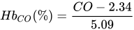
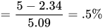
Carbon monoxide competes with oxygen for binding sites on hemoglobin molecules. As carbon monoxide binds with hemoglobin hundreds of times tighter than oxygen, it can prevent the carriage of oxygen.[69] Carbon monoxide poisoning can occur acutely, as with smoke intoxication, or over a period of time, as with cigarette smoking. Due to physiological processes, carbon monoxide is maintained at a resting level of 4–6 ppm. This is increased in urban areas (7–13 ppm) and in smokers (20–40 ppm).[70] A carbon monoxide level of 40 ppm is equivalent to a reduction in hemoglobin levels of 10 g/L.[70][note 1]
Carbon monoxide has another harmful effect: it changes how hemoglobin binds to oxygen, preventing the normal release of oxygen to body tissues and shifting the oxygen dissociation curve to the left, making the binding tighter. In so doing, the hemoglobin is less likely to release its oxygen at the peripheral tissues.[63] Certain abnormal hemoglobin variants also have higher than normal affinity for oxygen, and so are also poor at delivering oxygen to the periphery.[71]
Altitude
Atmospheric pressure reduces with altitude and proportionally, so does the oxygen content of the air.[72] The reduction in the partial pressure of inspired oxygen at higher altitudes lowers the oxygen saturation of the blood, ultimately leading to hypoxia.[72] The clinical features of altitude sickness include: sleep problems, dizziness, headache and oedema.[72]
Hypoxic breathing gases
The breathing gas may contain an insufficient partial pressure of oxygen. Such situations may lead to unconsciousness without symptoms since carbon dioxide levels remain normal and the human body senses pure hypoxia poorly. Hypoxic breathing gases can be defined as mixtures with a lower oxygen fraction than air, though gases containing sufficient oxygen to reliably maintain consciousness at normal sea level atmospheric pressure may be described as normoxic even when the oxygen fraction is slightly below normoxic. Hypoxic breathing gas mixtures in this context are those which will not reliably maintain consciousness at sea level pressure.[73]
One of the most widespread circumstances of exposure to hypoxic breathing gas is ascent to altitudes where the ambient pressure drops sufficiently to reduce the partial pressure of oxygen to hypoxic levels.[72]
Gases with as little as 2% oxygen by volume in a helium diluent are used for deep diving operations. The ambient pressure at 190 msw is sufficient to provide a partial pressure of about 0.4 bar, which is suitable for saturation diving. As the divers are decompressed, the breathing gas must be oxygenated to maintain a breathable atmosphere.[74]
It is also possible for the breathing gas for diving to have a dynamically controlled oxygen partial pressure, known as a set point, which is maintained in the breathing gas circuit of a diving rebreather by addition of oxygen and diluent gas to maintain the desired oxygen partial pressure at a safe level between hypoxic and hyperoxic at the ambient pressure due to the current depth. A malfunction of the control system may lead to the gas mixture becoming hypoxic at the current depth.[75]
A special case of hypoxic breathing gas is encountered in deep freediving where the partial pressure of the oxygen in the lung gas is depleted during the dive, but remains sufficient at depth, and when it drops during ascent, it becomes too hypoxic to maintain consciousness, and the diver loses consciousness before reaching the surface.[15][10]
Hypoxic gases may also occur in industrial, mining, and firefighting environments. Some of these may also be toxic or narcotic, others are just asphyxiant. Some are recognisable by smell, others are odourless.
Inert gas asphyxiation may be deliberate with use of a suicide bag. Accidental death has occurred in cases where concentrations of nitrogen in controlled atmospheres, or methane in mines, has not been detected or appreciated.[76]
Other
Hemoglobin function can also be impaired when the iron atom in its heme group is oxidized from the ferrous (Fe²⁺) to the ferric (Fe³⁺) state. This inactive form, known as methemoglobin, cannot bind oxygen effectively. Elevated levels of methemoglobin can occur following exposure to oxidizing substances such as sodium nitrite, nitrates, or certain medications[77]
Anemia
Hemoglobin plays a substantial role in carrying oxygen throughout the body,[63] and when it is deficient, anemia can result, causing 'anaemic hypoxia' if tissue oxygenation is decreased. Iron deficiency is the most common cause of anemia. As iron is used in the synthesis of hemoglobin, less hemoglobin will be synthesised when there is less iron, due to insufficient intake, or poor absorption.[64]:997–99
Anemia is typically a chronic process that is compensated over time by increased levels of red blood cells via upregulated erythropoetin. A chronic hypoxic state can result from a poorly compensated anaemia.[64]:997–99
Histotoxic hypoxia
Histotoxic hypoxia (also called histoxic hypoxia) is the inability of cells to take up or use oxygen from the bloodstream, despite physiologically normal delivery of oxygen to such cells and tissues.[78] Histotoxic hypoxia results from tissue poisoning, such as that caused by cyanide (which acts by inhibiting cytochrome oxidase) and certain other poisons like hydrogen sulfide (byproduct of sewage and used in leather tanning).[79]
Mechanism
Tissue hypoxia from low oxygen delivery may be due to low haemoglobin concentration (anaemic hypoxia), low cardiac output (stagnant hypoxia) or low haemoglobin saturation (hypoxic hypoxia).[80] The consequence of oxygen deprivation in tissues is a switch to anaerobic metabolism at the cellular level. As such, reduced systemic blood flow may result in increased serum lactate.[81] Serum lactate levels have been correlated with illness severity and mortality in critically ill adults and in ventilated neonates with respiratory distress.[81]
Physiological responses
All vertebrates must maintain oxygen homeostasis to survive, and have evolved physiological systems to ensure adequate oxygenation of all tissues. In air breathing vertebrates this is based on lungs to acquire the oxygen, hemoglobin in red corpuscles to transport it, a vasculature to distribute, and a heart to deliver. Short term variations in the levels of oxygenation are sensed by chemoreceptor cells which respond by activating existing proteins, and over longer terms by regulation of gene transcription. Hypoxia is also involved in the pathogenesis of some common and severe pathologies.[82]
The most common causes of death in an aging population include myocardial infarction, stroke and cancer. These diseases share a common feature that limitation of oxygen availability contributes to the development of the pathology. Cells and organisms are also able to respond adaptively to hypoxic conditions, in ways that help them to cope with these adverse conditions. Several systems can sense oxygen concentration and may respond with adaptations to acute and long-term hypoxia.[82] The systems activated by hypoxia usually help cells to survive and overcome the hypoxic conditions. Erythropoietin, which is produced in larger quantities by the kidneys under hypoxic conditions, is an essential hormone that stimulates production of red blood cells, which are the primary transporter of blood oxygen, and glycolytic enzymes are involved in anaerobic ATP formation.[51]
Hypoxia-inducible factors (HIFs) are transcription factors that respond to decreases in available oxygen in the cellular environment, or hypoxia.[83][84] The HIF signaling cascade mediates the effects of hypoxia on the cell. Hypoxia often keeps cells from differentiating. However, hypoxia promotes the formation of blood vessels, and is important for the formation of a vascular system in embryos and tumors. The hypoxia in wounds also promotes the migration of keratinocytes and the restoration of the epithelium.[85] It is therefore not surprising that HIF-1 modulation was identified as a promising treatment paradigm in wound healing.[86]
Exposure of a tissue to repeated short periods of hypoxia, between periods of normal oxygen levels, influences the tissue's later response to prolonged ischaemic exposure. This is known as ischemic preconditioning, and it is known to occur in many tissues.[51]
Acute
If oxygen delivery to cells is insufficient for the demand (hypoxia), electrons will be shifted to pyruvic acid in the process of lactic acid fermentation. This temporary measure (anaerobic metabolism) allows small amounts of energy to be released. Lactic acid buildup (in tissues and blood) is a sign of inadequate mitochondrial oxygenation, which may be due to hypoxemia, poor blood flow (e.g., shock) or a combination of both.[87] If severe or prolonged, it could lead to cell death.[88]
In humans, hypoxia is detected by the peripheral chemoreceptors in the carotid body and aortic body, with the carotid body chemoreceptors being the major mediators of reflex responses to hypoxia.[89] This response does not control ventilation rate at normal PO2, but below normal the activity of neurons innervating these receptors increases dramatically, so much as to override the signals from central chemoreceptors in the hypothalamus, increasing PO2 despite a falling PCO2[90]
In most tissues of the body, the response to hypoxia is vasodilation. By widening the blood vessels, the tissue allows greater perfusion.
By contrast, in the lungs, the response to hypoxia is vasoconstriction. This is known as hypoxic pulmonary vasoconstriction, or "HPV", and has the effect of redirecting blood away from poorly ventilated regions, which helps match perfusion to ventilation, giving a more even oxygenation of blood from different parts of the lungs.[82] In conditions of hypoxic breathing gas, such as at high altitude, HPV is generalized over the entire lung, but with sustained exposure to generalized hypoxia, HPV is suppressed.[91] Hypoxic ventilatory response (HVR) is the increase in ventilation induced by hypoxia that allows the body to take in and transport lower concentrations of oxygen at higher rates. It is initially elevated in lowlanders who travel to high altitude, but reduces significantly over time as people acclimatize.[4][92]
Chronic
Cuando la presión capilar pulmonar permanece elevada de forma crónica (durante al menos 2 semanas), los pulmones se vuelven aún más resistentes al edema pulmonar debido a que los vasos linfáticos se expanden considerablemente, aumentando su capacidad de evacuar el líquido de los espacios intersticiales hasta diez veces. Por lo tanto, en pacientes con estenosis mitral crónica , se han medido presiones capilares pulmonares de 40 a 45 mm Hg sin que se desarrolle edema pulmonar letal. [ 93 ]
Existen varios mecanismos fisiológicos potenciales para la hipoxemia, pero en pacientes con enfermedad pulmonar obstructiva crónica ( EPOC ), el desequilibrio ventilación/perfusión (V/Q) es el más común, con o sin hipoventilación alveolar, según lo indica la concentración arterial de dióxido de carbono. La hipoxemia causada por el desequilibrio V/Q en la EPOC es relativamente fácil de corregir, y se requieren flujos relativamente bajos de oxígeno suplementario (menos de 3 L/min para la mayoría de los pacientes) para la oxigenoterapia a largo plazo (OTLP). La hipoxemia normalmente estimula la ventilación y produce disnea, pero estos y otros signos y síntomas de hipoxia son suficientemente variables en la EPOC como para limitar su utilidad en la evaluación del paciente. La hipoxia alveolar crónica es el principal factor que conduce al desarrollo de cor pulmonale —hipertrofia ventricular derecha con o sin insuficiencia ventricular derecha manifiesta— en pacientes con EPOC. La hipertensión pulmonar afecta negativamente la supervivencia en la EPOC, proporcional a la elevación de la presión arterial pulmonar media en reposo. Aunque la gravedad de la obstrucción del flujo aéreo, medida mediante pruebas de volumen espiratorio forzado (VEF1) , se correlaciona mejor con el pronóstico general en la EPOC, la hipoxemia crónica aumenta la mortalidad y la morbilidad en cualquier grado de gravedad de la enfermedad. Estudios a gran escala sobre la oxigenoterapia a largo plazo en pacientes con EPOC muestran una relación dosis-respuesta entre las horas diarias de uso de oxígeno suplementario y la supervivencia. El uso continuo de oxígeno las 24 horas del día en pacientes seleccionados adecuadamente puede producir un beneficio significativo en la supervivencia. [ 6 ]
Respuestas patológicas
isquemia cerebral
The brain has relatively high energy requirements, using about 20% of the oxygen under resting conditions, but low reserves, which make it specially vulnerable to hypoxia. In normal conditions, an increased demand for oxygen is easily compensated by an increased cerebral blood flow. but under conditions when there is insufficient oxygen available, increased blood flow may not be sufficient to compensate, and hypoxia can result in brain injury. A longer duration of cerebral hypoxia will generally result in larger areas of the brain being affected. The brainstem, hippocampus and cerebral cortex seem to be the most vulnerable regions. Injury becomes irreversible if oxygenation is not soon restored. Most cell death is by necrosis but delayed apoptosis also occurs. In addition, presynaptic neurons release large amounts of glutamate which further increases Ca2+ influx and causes catastrophic collapse in postsynaptic cells. Although it is the only way to save the tissue, reperfusion also produces reactive oxygen species and inflammatory cell infiltration, which induces further cell death. If the hypoxia is not too severe, cells can suppress some of their functions, such as protein synthesis and spontaneous electrical activity, in a process called penumbra, which is reversible if the oxygen supply is resumed soon enough.[82]
Myocardial ischemia
Parts of the heart are exposed to ischemic hypoxia in the event of occlusion of a coronary artery. Short periods of ischaemia are reversible if reperfused within about 20 minutes, without development of necrosis, but the phenomenon known as stunning is generally evident. If hypoxia continues beyond this period, necrosis propagates through the myocardial tissue.[82] Energy metabolism in the affected area shifts from mitochondrial respiration to anaerobic glycolysis almost immediately, with concurrent reduction of effectiveness of contractions, which soon cease. Anaerobic products accumulate in the muscle cells, which develop acidosis and osmotic load leading to cellular edema. Intracellular Ca2+ increases and eventually leads to cell necrosis. Arterial flow must be restored to return to aerobic metabolism and prevent necrosis of the affected muscle cells, but this also causes further damage by reperfusion injury. Myocadial stunning has been described as "prolonged postischaemic dysfunction of viable tissue salvaged by reperfusion", which manifests as temporary contractile failure in oxygenated muscle tissue. This may be caused by a release of reactive oxygen species during the early stages of reperfusion.[82]
Tumor angiogenesis
A medida que los tumores crecen, se desarrollan regiones de hipoxia relativa debido a que las células tumorales utilizan el oxígeno de forma desigual. La formación de nuevos vasos sanguíneos es necesaria para el crecimiento continuo del tumor y también es un factor importante en la metástasis, ya que constituye la vía por la cual las células cancerosas se transportan a otros sitios. [ 82 ]
En condiciones de hipoxia, las células tumorales activan una respuesta molecular coordinada mediada principalmente por factores inducibles por hipoxia (HIF), en particular HIF-1α. La estabilización de HIF-1α conduce a la transcripción de numerosos genes proangiogénicos, incluidos el factor de crecimiento endotelial vascular (VEGF), el factor de crecimiento derivado de plaquetas (PDGF) y las angiopoyetinas , que promueven la proliferación y migración de células endoteliales, así como la formación de nuevos capilares alrededor del núcleo tumoral hipóxico. [ 94 ]
La vasculatura tumoral resultante suele presentar anomalías estructurales y funcionales, caracterizadas por diámetros vasculares irregulares, mayor permeabilidad y perfusión deficiente, lo que perpetúa la hipoxia local y la inestabilidad genómica. Este ciclo de retroalimentación potencia el potencial adaptativo del tumor y su resistencia a terapias como la radioterapia y la quimioterapia , las cuales dependen de una oxigenación adecuada para maximizar sus efectos citotóxicos. [ 95 ]
Además de promover la angiogénesis, la señalización de HIF también apoya la reprogramación metabólica hacia la glucólisis anaeróbica, mejora la expresión de metaloproteinasas de matriz (MMP) que degradan los componentes de la matriz extracelular y aumenta la expresión de moléculas de adhesión que facilitan la invasión tumoral y la diseminación metastásica. [ 96 ]
En conjunto, estos mecanismos convierten la angiogénesis inducida por hipoxia en un rasgo distintivo central de la progresión maligna y en un objetivo terapéutico importante en oncología. Actualmente se utilizan o se investigan agentes que bloquean la señalización del VEGF (como el bevacizumab ) o que inhiben la actividad del HIF-1α para contrarrestar la angiogénesis tumoral y mejorar los resultados del tratamiento. [ 97 ]
Diagnóstico
Examen físico e historial clínico
La hipoxia puede presentarse de forma aguda o crónica.
Acute presentation may include dyspnea (shortness of breath) and tachypnea (rapid, often shallow, breathing). Severity of symptom presentation is commonly an indication of severity of hypoxia. Tachycardia (rapid pulse) may develop to compensate for low arterial oxygen tension. Stridor may be heard in upper airway obstruction, and cyanosis may indicate severe hypoxia. Neurological symptoms and organ function deterioration occur when the oxygen delivery is severely compromised. In moderate hypoxia, restlessness, headache and confusion may occur, with coma and eventual death possible in severe cases.[8]
In chronic presentation, dyspnea following exertion is most commonly mentioned. Symptoms of the underlying condition that caused the hypoxia may be apparent, and can help with differential diagnosis. A productive cough and fever may be present with lung infection, and leg edema may suggest heart failure.[8]
Lung auscultation can provide useful information.[8]
Tests
An arterial blood gas test (ABG) may be done, which usually includes measurements of oxygen content, hemoglobin, oxygen saturation (how much of the hemoglobin is carrying oxygen), arterial partial pressure of oxygen (PaO2), partial pressure of carbon dioxide (PaCO2), blood pH level, and bicarbonate (HCO3)[98]
- An arterial oxygen tension (PaO2) less than 80 mmHg is considered abnormal, but must be considered in context of the clinical situation.[8]
- In addition to diagnosis of hypoxemia, the ABG may provide additional information, such as PCO2, which can help identify the etiology. The arterial partial pressure of carbon dioxide is an indirect measure of exchange of carbon dioxide with the air in the lungs, and is related to minute ventilation. PCO2 is raised in hypoventilation.[8]
- The normal range of PaO2:FiO2 ratio is 300 to 500 mmHg, if this ratio is lower than 300 it may indicate a deficit in gas exchange, which is particularly relevant for identifying acute respiratory distress syndrome (ARDS). A ratio of less than 200 indicates severe hypoxemia.[8]
- El gradiente alvéolo-arterial (Aa O 2 , [ 99 ] o gradiente A-a) es la diferencia entre la concentración alveolar (A) de oxígeno y la concentración arterial (a) de oxígeno. Es un parámetro útil para reducir el diagnóstico diferencial de la hipoxemia . [ 100 ] El gradiente A-a ayuda a evaluar la integridad de la unidad alvéolo-capilar. Por ejemplo, a gran altitud, la presión parcial de oxígeno arterial (Pa O 2 ) es baja, pero solo porque la presión parcial de oxígeno alveolar (PA O 2 ) también es baja. Sin embargo, en estados de desequilibrio ventilación-perfusión , como la embolia pulmonar o el cortocircuito de derecha a izquierda , el oxígeno no se transfiere eficazmente de los alvéolos a la sangre , lo que resulta en un gradiente Aa elevado. La Pa O 2 se puede obtener del análisis de gases en sangre arterial y la PA O 2 se calcula utilizando la ecuación de gases alveolares . [ 8 ]
- Un hematocrito anormalmente bajo (porcentaje de volumen de glóbulos rojos) puede indicar anemia.
Las radiografías o las tomografías computarizadas del tórax y las vías respiratorias pueden revelar anomalías que pueden afectar la ventilación o la perfusión. [ 101 ]
Una gammagrafía de ventilación/perfusión [ 102 ] , también llamada gammagrafía pulmonar V/Q, es un tipo de imagen médica que utiliza gammagrafía e isótopos médicos para evaluar la circulación de aire y sangre dentro de los pulmones de un paciente [ 103 ] [ 104 ] con el fin de determinar la relación ventilación/perfusión. La parte de ventilación de la prueba analiza la capacidad del aire para llegar a todas las partes de los pulmones, mientras que la parte de perfusión evalúa qué tan bien circula la sangre dentro de los pulmones.
Las pruebas de función pulmonar [ 101 ] pueden incluir:
- Pruebas que miden los niveles de oxígeno durante la noche [ 101 ]
- La prueba de caminata de seis minutos , que mide qué tan lejos puede caminar una persona en una superficie plana en seis minutos para evaluar la capacidad de ejercicio midiendo los niveles de oxígeno en respuesta al ejercicio. [ 101 ]
- Las mediciones diagnósticas que pueden ser relevantes incluyen: [ 105 ] Volúmenes pulmonares , incluyendo capacidad pulmonar, resistencia de las vías respiratorias , fuerza de los músculos respiratorios , capacidad de difusión.
- Otras pruebas de función pulmonar que pueden ser relevantes incluyen: [ 105 ] Espirometría , pletismografía corporal , técnica de oscilación forzada para calcular el volumen, la presión y el flujo de aire en los pulmones, respuesta a broncodilatadores , prueba de difusión de monóxido de carbono (DLCO), estudios de titulación de oxígeno , prueba de esfuerzo cardiopulmonar , broncoscopia y toracocentesis
Diagnóstico diferencial
El tratamiento dependerá de la gravedad y también puede depender de la causa, ya que algunos casos se deben a causas externas y eliminarlas y tratar los síntomas agudos puede ser suficiente, pero cuando los síntomas se deben a una patología subyacente, el tratamiento de los síntomas evidentes puede proporcionar solo un alivio temporal o parcial, por lo que el diagnóstico diferencial puede ser importante para seleccionar el tratamiento definitivo.
- Hipoxia hipoxémica : La baja tensión de oxígeno en la sangre arterial (PaO₂ ) generalmente indica la incapacidad de los pulmones para oxigenar la sangre adecuadamente. Las causas internas incluyen hipoventilación, difusión alveolar alterada y derivación pulmonar. Las causas externas incluyen un ambiente hipóxico, que podría ser causado por una baja presión ambiental o un gas respiratorio inadecuado. [ 8 ] Tanto la hipoxia aguda como la crónica y la hipercapnia causadas por disfunción respiratoria pueden producir síntomas neurológicos como encefalopatía, convulsiones, dolor de cabeza, papiledema y asterixis . [ 55 ] El síndrome de apnea obstructiva del sueño puede causar dolores de cabeza matutinos. [ 55 ]
- La hipoxia circulatoria se produce por una perfusión insuficiente de los tejidos afectados por sangre adecuadamente oxigenada. Esta puede ser generalizada, debido a insuficiencia cardíaca o hipovolemia, o localizada, debido a infarto o lesión localizada. [ 8 ]
- La hipoxia anémica es causada por un déficit en la capacidad de transporte de oxígeno, generalmente debido a niveles bajos de hemoglobina, lo que lleva a un suministro inadecuado generalizado de oxígeno. [ 8 ]
- La hipoxia histotóxica (disoxia) es consecuencia de la incapacidad de las células para utilizar el oxígeno de forma eficaz. Un ejemplo clásico es la intoxicación por cianuro, que inhibe la enzima citocromo c oxidasa en las mitocondrias, bloqueando el uso de oxígeno para producir ATP. [ 8 ]
Critical illness polyneuropathy or myopathy should be considered in the intensive care unit when patients have difficulty coming off the ventilator.[55]
Prevention
Prevention can be as simple as risk management of occupational exposure to hypoxic environments, and commonly involves the use of environmental monitoring and personal protective equipment. Prevention of hypoxia as a predictable consequence of medical conditions requires prevention of those conditions. Screening of demographics known to be at risk for specific disorders may be useful.
Prevention of altitude-induced hypoxia
To counter the effects of high-altitude diseases, the body must return arterial PaO2 toward normal. Acclimatization, the means by which the body adapts to higher altitudes, only partially restores PO2 to standard levels. Hyperventilation, the body's most common response to high-altitude conditions, increases alveolar PO2 by raising the depth and rate of breathing. However, while PO2 does improve with hyperventilation, it does not return to normal. Studies of miners and astronomers working at 3000 meters and above show improved alveolar PO2 with full acclimatization, yet the PO2 level remains equal to or even below the threshold for continuous oxygen therapy for patients with chronic obstructive pulmonary disease (COPD).[106] In addition, there are complications involved with acclimatization. Polycythemia, in which the body increases the number of red blood cells in circulation, thickens the blood, raising the risk of blood clots.[107]
En situaciones de gran altitud, solo el enriquecimiento de oxígeno o la presurización del compartimento pueden contrarrestar los efectos de la hipoxia. La presurización es factible en vehículos y para emergencias en instalaciones terrestres. Al aumentar la concentración de oxígeno a presión ambiente, se contrarrestan los efectos de la baja presión barométrica y se restablece el nivel de PO₂ arterial a su capacidad normal. Una pequeña cantidad de oxígeno suplementario reduce la altitud equivalente en habitaciones con climatización. A 4000 m, aumentar el nivel de concentración de oxígeno en un 5 % mediante un concentrador de oxígeno y un sistema de ventilación existente proporciona una altitud equivalente a 3000 m, lo que es mucho más tolerable para el creciente número de personas de tierras bajas que trabajan en altitudes elevadas. [ 108 ] En un estudio de astrónomos que trabajaban en Chile a 5050 m, los concentradores de oxígeno aumentaron el nivel de concentración de oxígeno en casi un 30 % (es decir, del 21 % al 27 %). Esto resultó en una mayor productividad de los trabajadores, menos fatiga y un mejor sueño. [ 106 ]
Los concentradores de oxígeno pueden utilizarse para el enriquecimiento de oxígeno a gran altitud en entornos con clima controlado . Requieren un mantenimiento mínimo, funcionan con un suministro de aire local y consumen relativamente poca electricidad. Al extraer el oxígeno directamente del aire ambiente, los concentradores eliminan la carga logística y financiera del transporte de cilindros de oxígeno comprimido a regiones remotas o montañosas. Dado que las oficinas y los edificios residenciales a gran altitud suelen mantener una temperatura y humedad controladas, estos sistemas pueden integrarse fácilmente en la infraestructura de climatización existente. [ 109 ]
Tratamiento y manejo
El tratamiento y el manejo dependen de las circunstancias. En la mayoría de las situaciones de gran altitud, el riesgo es conocido y la prevención es apropiada. A baja altitud, la hipoxia suele estar asociada a un problema médico o a una contingencia inesperada, y el tratamiento se adapta a cada caso específico. Es necesario identificar a las personas que requieren oxigenoterapia, ya que el oxígeno suplementario es necesario para tratar la mayoría de las causas de hipoxia, pero pueden ser apropiadas diferentes concentraciones de oxígeno. [ 110 ]
Tratamiento de casos agudos y crónicos
El tratamiento dependerá de la causa de la hipoxia. Si se determina que existe una causa externa y esta puede eliminarse, el tratamiento podría limitarse a brindar soporte y restablecer la oxigenación normal del organismo. En otros casos, podría ser necesario un tratamiento más prolongado, que podría requerir oxígeno suplementario durante un período considerable o de forma indefinida.
Existen tres aspectos principales del tratamiento de oxigenación: mantener las vías respiratorias permeables, proporcionar un contenido de oxígeno suficiente en el aire inspirado y mejorar la difusión en los pulmones. [ 8 ] En algunos casos, el tratamiento puede extenderse a la mejora de la capacidad de oxígeno en la sangre, lo que puede incluir intervención y soporte volumétrico y circulatorio, terapia de oxígeno hiperbárico y tratamiento de la intoxicación.
La ventilación invasiva puede ser necesaria o una opción electiva en cirugía. Generalmente , implica un ventilador de presión positiva conectado a un tubo endotraqueal, y permite una ventilación precisa, una monitorización exacta de la FiO₂ y la presión positiva al final de la espiración, y puede combinarse con la administración de gases anestésicos. En algunos casos, puede ser necesaria una traqueotomía . [ 8 ] La disminución de la tasa metabólica mediante la reducción de la temperatura corporal disminuye la demanda y el consumo de oxígeno, y puede minimizar los efectos de la hipoxia tisular, especialmente en el cerebro; la hipotermia terapéutica basada en este principio puede ser útil. [ 8 ]
Cuando el problema se debe a insuficiencia respiratoria, es conveniente tratar la causa subyacente. En casos de edema pulmonar, se pueden usar diuréticos para reducir el edema. Los esteroides pueden ser eficaces en algunos casos de enfermedad pulmonar intersticial y, en casos extremos, se puede utilizar la oxigenación por membrana extracorpórea (ECMO). [ 8 ]
Se ha demostrado que la oxigenoterapia hiperbárica es útil para tratar algunas formas de hipoxia localizada, incluidas las lesiones traumáticas con perfusión deficiente, como las lesiones por aplastamiento, el síndrome compartimental y otras isquemias traumáticas agudas. [ 111 ] [ 112 ] Es el tratamiento definitivo para la enfermedad por descompresión grave , que es principalmente una afección que implica hipoxia localizada causada inicialmente por embolia de gas inerte y reacciones inflamatorias al crecimiento de burbujas extravasculares. [ 113 ] [ 114 ] [ 115 ] También es eficaz en la intoxicación por monóxido de carbono [ 116 ] y el pie diabético . [ 117 ] [ 118 ]
La renovación de una receta de oxígeno domiciliario tras una hospitalización requiere una evaluación del paciente para detectar hipoxemia persistente. [ 119 ]
Resultados
Prognosis in cases of hypoxia depends strongly on the underlying cause, the severity and duration of oxygen deprivation, the effectiveness and timeliness of treatment, and any pre-existing medical conditions that impair oxygen delivery or utilization, such as anemia, COPD, or cardiovascular disease. Mild, short-term hypoxia is often reversible with prompt oxygen therapy and correction of the underlying cause, whereas prolonged or severe hypoxia can result in irreversible cellular injury, hypoxic brain injury, or multi-organ failure.[120]
Hypoxia that leads to impaired judgment, delayed reactions, or loss of consciousness can indirectly contribute to fatal outcomes in situations where the immediate cause of death is secondary. This has been documented in underwater diving accidents, where hypoxia can cause confusion or unconsciousness before drowning occurs; in high-altitude mountaineering, where it contributes to exposure, hypothermia, and falls; and in aviation, where pilots of unpressurized aircraft or performers in aerobatic maneuvers may lose control of the aircraft following hypoxic incapacitation.[121]
In occupational and environmental contexts, chronic or intermittent hypoxia—such as in miners, high-altitude workers, or individuals with sleep apnea—can have cumulative effects on cognitive performance, cardiovascular function, and metabolic regulation. Long-term exposure may increase the risk of pulmonary hypertension, polycythemia, and cardiac remodeling, though individual susceptibility varies.[122]
Epidemiology
Hypoxia is a common disorder but there are many possible causes.[8] Prevalence is variable. Some of the causes are very common, like pneumonia or chronic obstructive pulmonary disease; some are quite rare like hypoxia due to cyanide poisoning. Others, like reduced oxygen tension at high altitude, may be regionally distributed or associated with a specific demographic.[8]
Generalized hypoxia is an occupational hazard in several high-risk professions where oxygen availability is reduced or disrupted. These include firefighting, where smoke inhalation and exposure to carbon monoxide can impair oxygen transport; professional diving, where improper gas mixtures or equipment failure may lead to hypoxic hypoxia; mining and underground rescue, where oxygen depletion can result from combustion, gas displacement, or poor ventilation; and aviation, particularly when flying at high altitudes in unpressurized aircraft, where ambient oxygen pressure falls below levels needed for adequate blood oxygenation.[121]
Potentially life-threatening hypoxemia is common in critically ill patients.[123]
Localized hypoxia can occur when blood flow to a specific region of the body is restricted, leading to insufficient oxygen delivery to the affected tissues. This may arise as a complication of diabetes mellitus, where peripheral artery disease and microvascular damage impair circulation to the extremities; as a consequence of decompression sickness, in which gas emboli obstruct capillaries; or following traumatic injury that compromises local blood supply.[124][125]
Hypoxia related to underdeveloped lung function is also a frequent complication of premature birth. In preterm infants, incomplete maturation of the alveoli and insufficient production of surfactant can lead to respiratory distress syndrome, resulting in systemic oxygen deprivation.[126] In the United States, intrauterine hypoxia and birth asphyxia together were ranked as the tenth leading cause of infant mortality in recent national statistics.[127]
Silent hypoxia
Silent hypoxia (also known as happy hypoxia)[128][129] is generalised hypoxia that does not coincide with shortness of breath.[130][131][132] This presentation is known to be a complication of COVID-19,[133][134] and is also known in atypical pneumonia,[135] altitude sickness,[136][137][138] and rebreather malfunction accidents.[139][140]
History
The 2019 Nobel Prize in Physiology or Medicine was awarded to William G. Kaelin Jr., Sir Peter J. Ratcliffe, and Gregg L. Semenza in recognition of their discovery of cellular mechanisms to sense and adapt to different oxygen concentrations, establishing a basis for how oxygen levels affect physiological function.[141][142]
The use of the term hypoxia appears to be relatively recent, with the first recorded use in scientific publication from 1945. Previous to this the term anoxia was extensively used for all levels of oxygen deprivation. Investigation into the effects of lack of oxygen date from the mid 19th century. [143]
Etymology
Hypoxia is formed from the Greek roots υπo (hypo), meaning under, below, and less than, and oξυ (oxy), meaning acute or acid, which is the root for oxygen.[143]
See also
- Asphyxia – Severely deficient supply of oxygen
- Cerebral hypoxia – Oxygen shortage of the brain
- Erotic asphyxiation – Intentional restriction of oxygen to the brain for sexual arousal
- Fink effect, also known as diffusion hypoxia – Effect of anesthesia
- G-LOC – Loss of consciousness due to sustained high acceleration
- Histotoxic hypoxia – Medical condition in which cells cannot use oxygen
- Hyperoxia – Exposure of tissues to abnormally high concentrations of oxygen
- Hypoventilation training – Physical training method
- Hypoxemia – Abnormally low level of oxygen in the blood
- Hypoxia in fish – Response of fish to environmental hypoxia
- Factor inducible por hipoxia : proteína que responde a la baja concentración de oxígeno.
- Hipoxia hipóxica : condición médica de privación de oxígeno Páginas que muestran descripciones breves de objetivos de redirección , resultado de la insuficiencia de oxígeno disponible para los pulmones
- Respuesta ventilatoria hipóxica : reacción biológica al aumento de altitud.
- Hipoxificador – Dispositivo para suministrar aire respirable con contenido reducido de oxígeno; un dispositivo destinado a la aclimatación a la hipoxia de forma controlada.
- Entrenamiento hipóxico intermitente : técnica para mejorar el rendimiento humano mediante la adaptación a la reducción de oxígeno. Páginas que muestran breves descripciones de los objetivos de redireccionamiento.
- Hipoxia intrauterina : afección médica en la que el feto no recibe suficiente oxígeno.
- Hipoxia latente : concentración de oxígeno en los pulmones y en la sangre suficiente para mantener la conciencia solo a profundidad.
- Pseudohipoxia , aumento de la proporción citosólica de NADH libre a NAD + en las células
- Rinomanometría : método utilizado para evaluar la función respiratoria de la cavidad nasal.
- Apnea del sueño : trastorno que implica pausas en la respiración durante el sueño.
- Estrés sólido : una de las características físicas distintivas del cáncer.
- Tiempo de conciencia útil : duración del desempeño efectivo en un entorno hipóxico.
- Hipoxia tumoral : situación en la que las células tumorales han sido privadas de oxígeno.
- mimetismo vasculogénico
Notas
Referencias
- ↑ Samuel, Jacob; Franklin, Cory (2008). «Hipoxemia e hipoxia». Enfermedades quirúrgicas comunes . Nueva York: Springer. págs. 391–394 . doi : 10.1007/978-0-387-75246-4_97 . ISBN 978-0-387-75245-7.
- ↑ Das, Kusal K.; Honnutagi, Rajesh; Mullur, Lata; Reddy, R. Chandramouli; Das, Swastika; Majid, Dewan Syed Abdul; Biradar, MS (2019). "Metales pesados y microambiente con bajo contenido de oxígeno: su impacto en el metabolismo hepático y la suplementación dietética". Intervenciones dietéticas en la enfermedad hepática . págs. 315–332 . doi : 10.1016/B978-0-12-814466-4.00026-4 . ISBN 978-0-12-814466-4.
- ↑ West, John B. (1977). Fisiopatología pulmonar: Lo esencial . Williams & Wilkins. pág. 22. ISBN 978-0-683-08936-3.
- 1 2 Cymerman, A.; Rock, PB Problemas médicos en entornos de alta montaña. Un manual para oficiales médicos. Informe técnico USARIEM-TN94-2 (Informe). Instituto de Investigación de Medicina Ambiental del Ejército de los EE. UU. División de Medicina Térmica y de Montaña. DTIC ADA278095 .
- ↑ Gore, CJ; Clark, SA; Saunders, PU (septiembre de 2007). "Mecanismos no hematológicos de mejora del rendimiento a nivel del mar tras la exposición a la hipoxia" . Med Sci Sports Exerc . 39 (9): 1600–09 . doi : 10.1249/mss.0b013e3180de49d3 . PMID 17805094 .
- 1 2 Pierson, DJ (2000). "Fisiopatología y efectos clínicos de la hipoxia crónica". Respir Care . 45 (1): 39– 51, discusión 51–53. PMID 10771781 .
- 1 2 3 4 5 6 7 8 9 10 11 12 Complicaciones en Neuroanestesia . 2016. doi : 10.1016/C2015-0-00811-5 . ISBN 978-0-12-804075-1.
- 1 2 3 4 5 6 7 8 9 10 11 12 13 14 15 16 17 18 19 20 21 22 23 24 25 26 Bhutta, Beenish S.; Alghula, Faysal; Berim, Ilya (2025). "Hipoxia" . EstadísticasPerlas . Publicación de StatPearls. PMID 29493941 .
- ↑ Elliott, David (1997). "Algunas limitaciones de los rebreathers semicerrados". South Pacific Underwater Medicine Society Journal . 27 (1). ISSN 0813-1988 . OCLC 16986801 .
- 1 2 Lindholm, Peter (2006). Lindholm, P.; Pollock, NW; Lundgren, CEG (eds.). Mecanismos fisiológicos implicados en el riesgo de pérdida de conciencia durante el buceo en apnea (PDF) . Buceo en apnea. Actas del taller de la Sociedad de Medicina Submarina e Hiperbárica/Red de Alerta para Buzos, 20-21 de junio de 2006. Durham, NC: Red de Alerta para Buzos. pág. 26. ISBN 978-1-930536-36-4Consultado el 24 de enero de 2017 .
- 12"Hypoxia: Management and Treatment". my.clevelandclinic.org. Retrieved 27 November 2022.
- ↑Bleecker, M.L. (2015). "Carbon monoxide intoxication". Occupational Neurology. Handbook of Clinical Neurology. Vol. 131. pp. 191–203. doi:10.1016/B978-0-444-62627-1.00024-X. ISBN 978-0-444-62627-1. PMID 26563790.
- 12Mandal, Ananya (17 February 2010). "Hypoxia Types". www.news-medical.net. Retrieved 27 November 2022.
- ↑Levine, B.D. (2002). "Intermittent hypoxic training: fact and fancy". High Altitude Medicine & Biology. 3 (2): 177–93. doi:10.1089/15270290260131911. PMID 12162862.
- 12Pearn, John H.; Franklin, Richard C.; Peden, Amy E. (2015). "Hypoxic Blackout: Diagnosis, Risks, and Prevention". International Journal of Aquatic Research and Education. 9 (3): 342–347. doi:10.25035/ijare.09.03.09– via ScholarWorks@BGSU.
- ↑Della Rocca, Ylenia; Fonticoli, Luigia; Rajan, Thangavelu Soundara; Trubiani, Oriana; Caputi, Sergio; Diomede, Francesca; Pizzicannella, Jacopo; Marconi, Guya Diletta (2022). "Hypoxia: molecular pathophysiological mechanisms in human diseases". Journal of Physiology and Biochemistry. 78 (4): 739–752. doi:10.1007/s13105-022-00912-6. PMC 9684243. PMID 35870078.
- 12Robinson, Grace; Strading, John; West, Sophie (2009). Oxford Handbook of Respiratory Medicine. Oxford University Press. p. 880. ISBN 978-0-19-954516-2.
- ↑Zhou, Qiquan (June 1, 2011). "Standardization of Methods for Early Diagnosis and On-Site Treatment of High-Altitude Pulmonary Edema". Pulm Med. 2011 (7) 190648. doi:10.1155/2011/190648. PMC 3109313. PMID 21660284.
- ↑ Bergqvist, Pia (15 de abril de 2015). "Prevención de la hipoxia: ¿Qué hacer ahora?" . Flying . Archivado del original el 21 de septiembre de 2017. Recuperado el 22 de abril de 2015 .
- ↑ Illingworth, Robin; Graham, Colin; Hogg, Kerstin (2012). Oxford Handbook of Emergency Medicine . Oxford University Press. p. 768. ISBN 978-0-19-958956-2.
- ↑ Hillman, Ken; Bishop, Gillian (2004). Cuidados intensivos clínicos y medicina aguda . Cambridge University Press. pág. 685. ISBN 978-1-139-44936-6.
- ↑ Longmore, J.; Longmore, Murray; Wilkinson, Ian; Rajagopalan, Supraj (2006). Mini Oxford Handbook of Clinical Medicine . Oxford University Press. pág. 874. ISBN 978-0-19-857071-4.
- ↑ Ahrens, Thomas; Rutherford Basham, Kimberley (1993). Fundamentos de la oxigenación: Implicaciones para la práctica clínica . Jones & Bartlett Learning. pág. 194. ISBN 978-0-86720-332-5.
- ↑ Ramrakha, Punit; Moore, Kevin (2004). Oxford Handbook of Acute Medicine . Oxford University Press. p. 990. ISBN 978-0-19-852072-6.
- ↑ Wang, Bin; Zeng, Hongtao; Liu, Jingliu; Sun, Miao (25-10-2021). "Efectos de la hipoxia prenatal en el desarrollo del sistema nervioso y enfermedades relacionadas" . Frontiers in Neuroscience . 15 755554. doi : 10.3389/fnins.2021.755554 . PMC 8573102. PMID 34759794 .
- ↑ Choudhary, Mukesh; Sharma, Deepak; Dabi, Dhanraj; Lamba, Mamta; Pandita, Aakash; Shastri, Sweta (2015-01-12). " Disfunción hepática en neonatos asfixiados: estudio prospectivo de casos y controles" . Clinical Medicine Insights. Pediatrics . 9 CMPed.S21426: 1– 6. doi : 10.4137/CMPed.S21426 . PMC 4294631. PMID 25674030 .
- ↑ "Enfermedad arterial periférica oclusiva" . El manual de salud en el hogar de Merck . Merck & Co. Marzo de 2010. Consultado el 4 de marzo de 2012 .
- ↑ "Isquemia crónica que amenaza las extremidades (CLTI) – Vascular Cures" . vascularcures.org . Archivado del original el 29/10/2021 . Consultado el 27/10/2021 .
- ↑Zhai, Y; Petrowsky, H.; Hong, J.C.; Busuttil, R.W.; Kupiec-Weglinski, J.W. (10 February 2013). "Ischaemia–reperfusion injury in liver transplantation — From bench to bedside". Nat Rev Gastroenterol Hepatol. 10 (2): 79–89. doi:10.1038/nrgastro.2012.225. PMC 3577927. PMID 23229329.
- ↑Perico, N; Cattaneo, D.; Sayegh, M.H.; Remuzzi, G. (November 2004). "Delayed graft function in kidney transplantation". Lancet. 364 (9447): 1814–1827. doi:10.1016/S0140-6736(04)17406-0. PMID 15541456.
- 12Pmhdev. "Compartment Syndrome – National Library of Medicine". PubMed Health. Archived from the original on 10 September 2017. Retrieved 25 July 2017.
- ↑Peitzman, Andrew B.; Rhodesn, Michael; Schwab, C. William (2008). The Trauma Manual: Trauma and Acute Care Surgery. Lippincott Williams & Wilkins. p. 349. ISBN 978-0-7817-6275-5. Archived from the original on 2017-07-29.
- ↑Ferri, Fred F. (2017). Ferri's Clinical Advisor 2018 E-Book: 5 Books in 1. Elsevier Health Sciences. p. 317. ISBN 978-0-323-52957-0. Archived from the original on 2017-07-29.
- ↑Mascarenhas, Alexander; Braga, Alice; Majernikova, Sara Maria; Nizari, Shereen; Marletta, Debora; Theparambil, Shefeeq M.; Aziz, Qadeer; Marina, Nephtali; Gourine, Alexander V. (2025). "On the mechanisms of brain blood flow regulation during hypoxia". The Journal of Physiology. 603 (8): 2263–2280. doi:10.1113/JP285060. ISSN 1469-7793. PMC 12013793. PMID 38843467.
- ↑Malhotra, R. (November 2001). "Hypoxia induces apoptosis via two independent pathways in Jurkat cells: differential regulation by glucose". American Journal of Physiology. Cell Physiology. 281 (5): C1596–C1603. doi:10.1152/ajpcell.2001.281.5.c1596. PMID 11600423.
- ↑ Mattiesen, WR; et al. (mayo de 2009). "El aumento de la neurogénesis tras la encefalopatía hipóxico-isquémica en humanos está relacionado con la edad" . Acta Neuropathol . 117 (5): 525– 534. doi : 10.1007/s00401-009-0509-0 . PMID 19277687 .
- ↑ Robinson, LR; Micklesen, PJ; Tirschwell, DL; Lew, HL (marzo de 2003). "Valor predictivo de los potenciales evocados somatosensoriales para el despertar del coma". Critical Care Medicine . 31 (3): 960– 967. doi : 10.1097/01.ccm.0000053643.21751.3b . PMID 12627012 .
- ↑ Geraghty, MC; Torbey, MT (2006). "Neuroimágenes y marcadores serológicos de lesión neurológica después de un paro cardíaco". Neurol Clin . 24 (1): 107– 121. doi : 10.1016/j.ncl.2005.10.006 . PMID 6443133 .
- ↑ Busl, KM; Greer, DM (enero de 2010). "Lesión cerebral hipóxico-isquémica: fisiopatología, neuropatología y mecanismos" . NeuroRehabilitation . 26 (1): 5–13 . doi : 10.3233/NRE-2010-0531 . PMID 20130351 .
- 1 2 Condiciones, Grupo de Trabajo del Consejo Nacional de Investigación (EE. UU.) sobre el uso de lentes de contacto en condiciones adversas; Flattau, Pamela Ebert (1991), "Hipoxia" , Consideraciones sobre el uso de lentes de contacto en condiciones adversas: Actas de un simposio , National Academies Press (EE. UU.) , consultado el 11 de junio de 2023
- ↑ Tarvonen, M.; Hovi, P.; Sainio, S.; Vuorela, P.; Andersson, S.; Teramo, K. (2021). " Patrones cardiotocográficos intraparto y resultados perinatales relacionados con la hipoxia en embarazos complicados por diabetes mellitus gestacional" . Acta Diabetologica . 58 (11): 1563– 1573. doi : 10.1007/s00592-021-01756-0 . PMC 8505288. PMID 34151398 .
- ↑ Maslova, MV; Maklakova, AS; Sokolova, NA; Ashmarin, IP; Goncharenko, EN; Krushinskaya, YV (julio de 2003). "Los efectos de la hipoxia prenatal y postnatal en el sistema nervioso central y su corrección con hormonas peptídicas". Neuroscience and Behavioral Physiology . 33 (6): 607–11 . doi : 10.1023/A:1023938905744 . PMID 14552554 .
- ^ Habek, D.; Habek, JC; Jugović, D.; Salihagić, A. (2002). "Intrauterina hipoksija i sindrom iznenadne dojenačke smrti" [ Hipoxia intrauterina y síndrome de muerte súbita del lactante ] . Acta Medica Croatica (en croata). 56 (3): 109–18 . PMID 12630342 .
- ↑ Peleg, D.; Kennedy, CM; Hunter, SK (agosto de 1998). "Restricción del crecimiento intrauterino: identificación y manejo". American Family Physician . 58 (2): 453–60 , 466–7 . PMID 9713399 .
- ↑ Rosenberg, A. (junio de 2008). "El recién nacido con RCIU". Seminars in Perinatology . 32 (3): 219– 24. doi : 10.1053/j.semperi.2007.11.003 . PMID 18482625 .
- ↑ Gonzalez, FF; Miller, SP (noviembre de 2006). "¿La asfixia perinatal afecta la función cognitiva sin parálisis cerebral?" . Archives of Disease in Childhood. Fetal and Neonatal Edition . 91 (6): F454-9. doi : 10.1136/adc.2005.092445 . PMC 2672766 . PMID 17056843 .
- ↑ Bulterys, MG; Greenland, S.; Kraus, JF (octubre de 1990). "Hipoxia fetal crónica y síndrome de muerte súbita del lactante: interacción entre el tabaquismo materno y el hematocrito bajo durante el embarazo". Pediatrics . 86 (4): 535– 40. doi : 10.1542/peds.86.4.535 . PMID 2216618 .
- ^ Emami Nejad, Asiah; Najafgholian, Simin; Rostami, Alireza; Sistani, Alireza; Shojaeifar, Samaneh; Esparvarinha, Mojgán; Nedaeinia, Reza; Haghjooy Javanmard, Shaghayegh; Taherian, Marjan; Ahmadlou, Mojtaba; Salehi, Rasoul; Sadeghi, Bahmán; Manian, Mostafa (2021). "El papel de la hipoxia en el microambiente tumoral y el desarrollo de células madre cancerosas: un enfoque novedoso para desarrollar un tratamiento" . Cáncer Cell Internacional . 21 (1) 62.doi : 10.1186 /s12935-020-01719-5 . PMC 7816485 . PMID 33472628 .
- ↑ Gilkes, DM; Semenza, GL; Wirtz, D. (junio de 2014). "Hipoxia y matriz extracelular: impulsores de la metástasis tumoral" . Nature Reviews. Cancer . 14 (6): 430– 9. Bibcode : 2014NatRC..14..430G . doi : 10.1038/nrc3726 . PMC 4283800. PMID 24827502 .
- ↑ Spill, F.; Reynolds, DS; Kamm, RD; Zaman, MH (agosto de 2016). "Impacto del microambiente físico en la progresión tumoral y la metástasis" . Current Opinion in Biotechnology . 40 : 41–48 . doi : 10.1016/j.copbio.2016.02.007 . PMC 4975620. PMID 26938687 .
- 1 2 3 Lumb, Andrew B. (2017). Fisiología respiratoria aplicada de Nunn (8.ª ed.). Elsevier. ISBN 978-0-7020-6294-0.
- ↑ Urbani, Luca; Porcù, Silvio; De Angelis, Claudio; Farrace, Stefano; Antonini, Riccardo (1994). "Movimientos oculares de seguimiento suave durante y después de la exposición aguda a hipoxia hipobárica". En d'Ydewalle, Géry; Van Rensbergen, Johan (eds.). Funciones visuales y oculomotoras: avances en la investigación de los movimientos oculares . Estudios en procesamiento de información visual. Vol. 5. Elsevier. pp. 133–143 . doi : 10.1016/B978-0-444-81808-9.50018-4 . ISBN 978-0-444-81808-9.
- 1 2 3 4 5 6 7 8 9 10 11 Mandal, Ananya (17 de febrero de 2010). "Síntomas de hipoxia" . www.news-medical.net . Recuperado el 27 de noviembre de 2022 .
- 12345"17: Aeromedical Factors". Pilot's Handbook of Aeronautical Knowledge: FAA Manual H-8083-25. Washington, DC: Flight Standards Service. Federal Aviation Administration, U.S. Dept. of Transportation. 2001. ISBN 1-56027-540-5.
- 123456Prasad, Shweta; Pal, Pramod Kumar; Chen, Robert (2021). "Breathing and the Nervous System". Aminoff's Neurology and General Medicine. pp. 3–19. doi:10.1016/B978-0-12-819306-8.00001-0. ISBN 978-0-12-819306-8.
- ↑"A Quick Look at Reflexes - Health Encyclopedia - University of Rochester Medical Center".
- ↑Vingrys, A.J.; Garner, L.F. (June 1987). "The effect of a moderate level of hypoxia on human color vision". Doc Ophthalmol. 66 (2): 171–85. doi:10.1007/BF00140454. PMID 3691296.
- ↑Kam, C.A.; Yoong, Florence F.Y.; Ganendran, A. (1978). "Cortical blindness following hypoxia during cardiac arrest". Anaesth. Intens. Care. 6 (2): 143–145. doi:10.1177/0310057X7800600209. PMID 665992.
- ↑Kenneth Baillie; Alistair Simpson. "Altitude oxygen calculator". Apex (Altitude Physiology Expeditions). Archived from the original on 2017-06-11. Retrieved 2006-08-10. – Online interactive oxygen delivery calculator.
- ↑Patel, Sagar; Jose, Alvin; Mohiuddin, Shamim S. (2024), "Physiology, Oxygen Transport And Carbon Dioxide Dissociation Curve", StatPearls, Treasure Island (FL): StatPearls Publishing, PMID 30969637, retrieved 2024-08-23
- ↑Jae-Hwan, Lee; Eun-Kyeong, Shin; Dongoh, Lee; Eui-Bae, Jeung (April 2015). "SAT-532: Expression of Beta-Oxidation Related Genes Under Hypoxic Condition Induced Preeclamptic Model in Vitro and in Vivo". Endocrine Reviews. 36 (2).
- ↑ Collins, Julie-Ann; Rudenski, Aram; Gibson, John; Howard, Luke; O'Driscoll, Ronan (septiembre de 2015). " Relación entre la presión parcial de oxígeno, la saturación y el contenido: la curva de disociación de la hemoglobina-oxígeno" . Breathe . 11 (3): 194–201 . doi : 10.1183/20734735.001415 . PMC 4666443. PMID 26632351 .
- 1 2 3 Martin, Lawrence (1999). Todo lo que realmente necesitas saber para interpretar los gases arteriales (2.ª ed.). Filadelfia: Lippincott Williams & Wilkins. ISBN 978-0-683-30604-0.
- 1 2 3 Colledge, Nicki R.; Walker, Brian R.; Ralston, Stuart H., eds. (2010). Principios y práctica de la medicina de Davidson . Ilustrado por Robert Britton (21.ª ed.). Edimburgo: Churchill Livingstone/Elsevier. ISBN 978-0-7020-3085-7.
- ↑ Levin, Marvin E.; O'Neal, Lawrence W.; Bowker, John H. (1993). El pie diabético . Mosby Year Book. ISBN 978-0-8016-6878-4.
- ↑ Basbaum, Allan I.; Bautista, Diana M.; Scherrer, Grégory; Julius, David (2009). "Mecanismos celulares y moleculares del dolor" . Cell . 139 ( 2): 267– 284. doi : 10.1016/j.cell.2009.09.028 . PMC 2852643. PMID 19837031 .
- ↑ Burton, RR (1988). "Pérdida de conciencia inducida por G: definición, historia, estado actual". Aviation, Space, and Environmental Medicine . 59 (1): 2– 5. PMID 3281645 .
- ↑ West, John B. (2008). Fisiología respiratoria: Lo esencial (8.ª ed.). La Jolla: Wolters Kluwer Lippincott Williams & Wilkins. págs. 88–89 . ISBN 978-0-7817-7206-8.
- ↑ Douglas, CG; Haldane, JS; Haldane, JB (12 de junio de 1912). "Las leyes de combinación de la hemoglobina con monóxido de carbono y oxígeno" . The Journal of Physiology . 44 (4): 275– 304. Bibcode : 1912JPhsg..44..275D . doi : 10.1113/jphysiol.1912.sp001517 . PMC 1512793. PMID 16993128 .
- 1 2 3 Wald, NJ; Idle, M.; Boreham, J.; Bailey, A. (1981). "Monóxido de carbono en el aliento en relación con el tabaquismo y los niveles de carboxihemoglobina" . Thorax . 36 ( 5): 366– 369. doi : 10.1136/thx.36.5.366 . PMC 471511. PMID 7314006 .
- ↑ Finch, Stuart C. (1986). "Reseña del libro Hemoglobina: aspectos moleculares, genéticos y clínicos". New England Journal of Medicine . 315 (18): 1175. doi : 10.1056/NEJM198610303151831 .
- 1 2 3 4 Netzer, Nikolaus; Strohl, Kingman; Faulhaber, Martín; Gatterer, Hannes; Burtscher, Martin (1 de julio de 2013). "Enfermedades de las alturas relacionadas con la hipoxia" . Revista de medicina de viajes . 20 (4): 247– 55. doi : 10.1111/jtm.12017 . PMID 23809076 .
- ↑ Hausserman, Georgina (1 de mayo de 2017). "Gases respiratorios" . Divers Alert Network . Consultado el 2 de diciembre de 2022 .
- ↑ "15". Manual de buceo de la Armada de los EE. UU., 6.ª revisión . Estados Unidos: Comando de Sistemas Navales de los EE. UU. 2006. Archivado del original el 2 de mayo de 2008. Recuperado el 15 de junio de 2008 .
- ↑ Parker, Martin (noviembre de 2012). "Manual de usuario del rebreather" (PDF) . apdiving.com . Ambient Pressure Diving Ltd. Recuperado el 11 de mayo de 2021 .
- ↑ Milroy, Christopher (otoño de 2018). " Muertes por hipoxia ambiental y dióxido de carbono elevado" . Academic Forensic Pathology . 8n (1): 2– 7. doi : 10.23907/2018.001 . PMC 6474450. PMID 31240022 .
- ↑ Percy, Melanie J.; Lappin, Terry R. (2008). "Metahemoglobinemia congénita recesiva: deficiencia de citocromo b 5 reductasa". British Journal of Haematology . 141 (3): 298– 308. doi : 10.1111/j.1365-2141.2008.07017.x . PMID 18318771 .
- ↑"Forms of hypoxia". courses.kcumb.edu. Archived from the original on 2007-12-22.
- ↑Pittman, R.N. (2011). "7". Regulation of Tissue Oxygenation. Morgan & Claypool Life Sciences.
- ↑Lacroix, Jacques; Tucci, Marisa; Tinmouth, Alan; Gauvin, France; Karam, Oliver (2011). Pediatric Critical Care. Elsevier. pp. 1162–76. doi:10.1016/b978-0-323-07307-3.10082-5. ISBN 978-0-323-07307-3.
- 12Kluckow, Martin; Seri, Istvan (2012). Hemodynamics and Cardiology: Neonatology Questions and Controversies. Elsevier. pp. 237–67. doi:10.1016/b978-1-4377-2763-0.00012-3. ISBN 978-1-4377-2763-0.
- 1234567Michiels, Carine. (June 2004). "Physiological and pathological responses to hypoxia". American Journal of Pathology. 164 (6). Elsevier: 1875–1882. doi:10.1016/S0002-9440(10)63747-9. PMC 1615763. PMID 15161623.
- ↑Smith, T.G.; Robbins, P.A.; Ratcliffe, P.J. (May 2008). "The human side of hypoxia-inducible factor". British Journal of Haematology. 141 (3): 325–34. doi:10.1111/j.1365-2141.2008.07029.x. PMC 2408651. PMID 18410568.
- ↑Wilkins, S.E.; Abboud, M.I.; Hancock, R.L.; Schofield, C.J. (April 2016). "Targeting Protein-Protein Interactions in the HIF System". ChemMedChem. 11 (8): 773–86. doi:10.1002/cmdc.201600012. PMC 4848768. PMID 26997519.
- ↑Benizri, E.; Ginouvès, A.; Berra, E. (April 2008). "The magic of the hypoxia-signaling cascade". Cellular and Molecular Life Sciences. 65 (7–8): 1133–49. doi:10.1007/s00018-008-7472-0. PMC 11131810. PMID 18202826.
- ↑ Duscher, D.; Januszyk, M.; Maan, ZN; Whittam, AJ; Hu, MS; Walmsley, GG; Dong, Y.; Khong, SM; Longaker, MT; Gurtner, GC (marzo de 2017). "Comparación del inhibidor de la hidroxilasa dimetiloxalilglicina y el quelante de hierro deferoxamina en la cicatrización de heridas diabéticas y envejecidas" . Cirugía plástica y reconstructiva . 139 (3): 695e– 706e. doi : 10.1097/PRS.0000000000003072 . PMC 5327844. PMID 28234841 .
- ↑ Hobler, KE; Carey, LC (1973). " Efecto de la hipoxemia progresiva aguda sobre el gasto cardíaco y el exceso de lactato plasmático" . Ann Surg . 177 (2): 199– 202. doi : 10.1097/00000658-197302000-00013 . PMC 1355564. PMID 4572785 .
- ↑ Fulda, Simone; Gorman, Adrienne M.; Hori, Osamu; Samali, Afshin (2010). "Respuestas al estrés celular: supervivencia y muerte celular" . International Journal of Cell Biology . 2010 214074. doi : 10.1155/2010/214074 . PMC 2825543. PMID 20182529 .
- ↑ Arieff, Allen I. (2013). Hipoxia, acidosis metabólica y circulación . Springer. págs. 4–5 . ISBN 978-1-4614-7542-2.
- ↑ Prabhakar, Nanduri R.; Peers, Chris (2014). "Regulación de los canales iónicos por gasotransmisores: un paso clave en la detección de O 2 por el cuerpo carotídeo" . Fisiología . 29 (1): 49– 57. doi : 10.1152/physiol.00034.2013 . PMC 3929115. PMID 24382871 .
- ^ Gao, Yuansheng; Kuebler, Wolfgang M.; Raj, J. Usha (2021). "Biología vascular". En Broaddus, V. Courtney; Ernst, Joel; Rey, Jr, Talmadge E.; Lázaro, Esteban; Sarmiento, Kathleen F.; Schnapp, Lynn M.; Stapleton, Renée; Gotway, Michael B. (eds.). Libro de texto de medicina respiratoria de Murray y Nadel (7ª ed.). Elsevier. págs. 76 a 87. ISBN 978-0-323-65587-3.
- ↑ Teppema, Luc J.; Dahan, Albert (2010). "La respuesta ventilatoria a la hipoxia en mamíferos: mecanismos, medición y análisis". Physiological Reviews . 90 (2): 675– 754. doi : 10.1152/physrev.00012.2009 . PMID 20393196 .
- ↑ Hall, John E. (20 de mayo de 2015). Guyton and Hall Textbook of Medical Physiology (13.ª ed.). Saunders. ISBN 978-1-4557-7005-2.
- ↑ Semenza, Gregg L. (2003). "Dirigiendo la terapia contra el cáncer hacia HIF-1". Nature Reviews Cancer . 3 (10): 721– 732. doi : 10.1038/nrc1187 . PMID 13130303 .
- ↑ Carmeliet, Peter; Jain, Rakesh K. (2011). "Principios y mecanismos de normalización vascular para el cáncer y otras enfermedades angiogénicas". Nature Reviews Drug Discovery . 10 (6): 417– 427. Bibcode : 2011NRvDD..10..417C . doi : 10.1038/nrd3455 . PMID 21629292 .
- ↑ Rankin, Erinn B.; Giaccia, Amato J. (2016). "Control hipóxico de la metástasis" . Science . 352 ( 6282): 175– 180. Bibcode : 2016Sci...352..175R . doi : 10.1126/science.aaf4405 . PMC 4898055. PMID 27124451 .
- ↑ Hickey, Michele M.; Simon, M. Celeste (2006). Regulación de la angiogénesis por hipoxia y factores inducibles por hipoxia . Temas actuales en biología del desarrollo. Vol. 76. págs. 217–257 . doi : 10.1016/S0070-2153(06)76007-0 . ISBN 978-0-12-153176-8. PMID 17118268 .
- ↑ "Gases en sangre arterial (GSA)" . my.clevelandclinic.org . Consultado el 3 de diciembre de 2022 .
- ↑ Logan, Carolynn M.; Rice, M. Katherine (1987). Abreviaturas médicas y científicas de Logan . Filadelfia: JB Lippincott Company . pág . 4. ISBN 0-397-54589-4.
- ↑ Hantzidiamantis, PJ; Amaro, E. (2020). Fisiología, gradiente de oxígeno alveolar a arterial . StatPearls. PMID 31424737. NBK545153.
- 1 2 3 4 "Diagnóstico" . www.medicalnewstoday.com . Consultado el 4 de diciembre de 2020 .
- ↑ "Diagnóstico y pruebas" . my.clevelandclinic.org . Consultado el 4 de diciembre de 2022 .
- ↑ "Gammagrafía de ventilación/perfusión pulmonar" . Centro Médico de la Universidad de Maryland . Consultado el 3 de enero de 2018 .
- ↑ Mortensen, Jann; Berg, Ronan MG (1 de enero de 2019). "Gammagrafía pulmonar en EPOC" . Seminars in Nuclear Medicine . 49 (1): 16– 21. doi : 10.1053/j.semnuclmed.2018.10.010 . PMID 30545511 .
- 1 2 "Pruebas de función pulmonar" . www.templehealth.org . Consultado el 4 de diciembre de 2022 .
- 1 2 West, John B.; Colegio Estadounidense de Médicos; Sociedad Estadounidense de Fisiología (2004). "La base fisiológica de las enfermedades de gran altitud". Anales de Medicina Interna . 141 (10): 789– 800. doi : 10.7326/0003-4819-141-10-200411160-00010 . PMID 15545679 .
- ↑ "Policitemia" . www.nhs.uk. 23 de octubre de 2017. Consultado el 10 de diciembre de 2022 .
- ↑ West, John B. (1995). "Enriquecimiento de oxígeno del aire ambiente para aliviar la hipoxia de gran altitud". Fisiología de la respiración . 99 (2): 225– 32. doi : 10.1016/0034-5687(94)00094-G . PMID 7777705 .
- ↑ Smith, KR, & Brown, L. (2018). "Estrategias de enriquecimiento de oxígeno para la habitabilidad en altitudes elevadas." Journal of Environmental Engineering, 144(7), 04018055.
- ↑ Wagstaff, Adrian J. (2014). "Oxigenoterapia". Oh's Intensive Care Manual . pp. 327–340.e3. doi : 10.1016/B978-0-7020-4762-6.00028-X . ISBN 978-0-7020-4762-6.
- ↑ Sociedad de Medicina Submarina e Hiperbárica. "Lesión por aplastamiento, síndrome compartimental y otras isquemias traumáticas agudas" . Consultado el 21 de agosto de 2011 .
- ↑Bouachour, G.; Cronier, P.; Gouello, J.P.; Toulemonde, J.L.; Talha, A; Alquier, P. (August 1996). "Hyperbaric oxygen therapy in the management of crush injuries: a randomized double-blind placebo-controlled clinical trial". The Journal of Trauma. 41 (2): 333–39. doi:10.1097/00005373-199608000-00023. PMID 8760546.
- ↑Undersea and Hyperbaric Medical Society. "Decompression Sickness or Illness and Arterial Gas Embolism". Retrieved 2011-08-21.
- ↑Brubakk, A.O.; Neuman, T.S., eds. (2003). Bennett and Elliott's physiology and medicine of diving (5th Rev ed.). United States: Saunders Ltd. p. 800. ISBN 978-0-7020-2571-6.
- ↑C., Acott (1999). "A brief history of diving and decompression illness". South Pacific Underwater Medicine Society Journal. 29 (2). ISSN 0813-1988. OCLC 16986801.
- ↑Piantadosi, C.A. (2004). "Carbon monoxide poisoning". Undersea & Hyperbaric Medicine. 31 (1): 167–77. PMID 15233173.
- ↑Undersea and Hyperbaric Medical Society. "Enhancement of Healing in Selected Problem Wounds". Retrieved 2011-08-21.
- ↑Zamboni, W.A.; Wong, H.P.; Stephenson, L.L.; Pfeifer, M.A. (September 1997). "Evaluation of hyperbaric oxygen for diabetic wounds: a prospective study". Undersea & Hyperbaric Medicine. 24 (3): 175–79. PMID 9308140.
- ↑American College of Chest Physicians; American Thoracic Society (September 2013), "Five Things Physicians and Patients Should Question", Choosing Wisely: an initiative of the ABIM Foundation, American College of Chest Physicians and American Thoracic Society, archived from the original on 3 November 2013, retrieved 6 January 2013
- ↑Lumb, A. B. (2016). Nunn’s Applied Respiratory Physiology, 8th ed. Elsevier.
- 12West, J. B. (2018). High-Altitude Medicine and Physiology, 6th ed. CRC Press.
- ↑ Eckardt, K.-U.; Kurtz, A. (2005). "Regulación de la producción de eritropoyetina". European Journal of Clinical Investigation . 35 : 13–19 . doi : 10.1111/j.1365-2362.2005.01525.x . PMID 16281953 .
- ↑ Grupo de Ensayo SRLF (2018). " Hipoxemia en la UCI: prevalencia, tratamiento y resultados" . Annals of Intensive Care . 8 (1) 82. doi : 10.1186/s13613-018-0424-4 . PMC 6089859. PMID 30105416 .
- ↑ Catrina, Sergiu-Bogdan; Zheng, Xiaowei (2021). "Hipoxia y factores inducibles por hipoxia en la diabetes y sus complicaciones" . Diabetologia . 64 ( 4): 709– 716. doi : 10.1007/S00125-021-05380-Z . PMC 7940280. PMID 33496820 .
- ↑ Vann, Richard D.; Butler, Frank K.; Mitchell, Simon J.; Moon, Richard E. (2011). "Enfermedad por descompresión". The Lancet . 377 (9760): 153– 164. Bibcode : 2011Lanc..377..153V . doi : 10.1016/S0140-6736(10)61085-9 . PMID 21215883 .
- ↑ Jobe, Alan H.; Bancalari, Eduardo (2001). "Displasia broncopulmonar". American Journal of Respiratory and Critical Care Medicine . 163 (7): 1723– 1729. doi : 10.1164/ajrccm.163.7.2011060 . PMID 11401896 .
- ↑ Centros para el Control y la Prevención de Enfermedades (CDC). «Estadísticas de mortalidad infantil del período 2021: Archivo de nacimientos y defunciones infantiles vinculados». Informes Nacionales de Estadísticas Vitales, vol. 72, n.º 4, 2023.
- ↑ Tobin, MJ; Laghi, F.; Jubran, A. (agosto de 2020). "Por qué la hipoxemia silenciosa por COVID-19 desconcierta a los médicos" . American Journal of Respiratory and Critical Care Medicine . 202 (3): 356– 360. doi : 10.1164/rccm.202006-2157CP . PMC 7397783. PMID 32539537 .
- ↑ LaMotte, S (7 de mayo de 2020). "Hipoxia silenciosa: pacientes con Covid-19 que deberían estar jadeando por aire pero no lo están" . CNN .
- ^ Pappas, S. (23 de abril de 2020). "'Silent hypoxia' may be killing COVID-19 patients. But there's hope". Live Science.
- ↑"Three reasons why COVID-19 can cause silent hypoxia". ScienceDaily. 19 November 2020.
- ↑Emily, H. (3 June 2020). "Silent hypoxia and its role in COVID-19 detection". News Medical.
- ↑Chandra, A.; Chakraborty, U.; Pal, J.; Karmakar, P. (September 2020). "Silent hypoxia: a frequently overlooked clinical entity in patients with COVID-19". BMJ Case Reports. 13 (9) e237207. doi:10.1136/bcr-2020-237207. PMC 7478026. PMID 32900744.
- ↑Levitan, R. (20 April 2020). "The Infection That's Silently Killing Coronavirus Patients". The New York Times.
- ↑Bowden, O. (12 May 2020). "What is 'silent hypoxia'? The coronavirus symptom patients don't know they have". Global News.
- ↑Ottestad, W. (2020). "COVID-19 patients with respiratory failure: what can we learn from aviation medicine?". British Journal of Anaesthesia. 125 (3): e280–e281. doi:10.1016/j.bja.2020.04.012. PMC 7165289. PMID 32362340.
- ↑Gillespie, C. "'Silent Hypoxia' Is Making Some Coronavirus Patients Critically Ill—Here's Why It's So Dangerous". Health.
- ↑Blanchet, D.; Greene, S. (12 July 2020). "Your Captain Speaking: Silent Hypoxia and COVID-19". EMS World.
- ↑"Rebreathers guide for beginners". apdiving.com. Retrieved 11 May 2021.
- ↑Sellers, Steven H. (2016). "An Overview of Rebreathers in Scientific Diving 1998–2013". In Pollock, N.W.; Sellers, S.H.; Godfrey, J.M. (eds.). Rebreathers and Scientific Diving(PDF). Proceedings of NPS/NOAA/DAN/AAUS Workshop, 16–19 June 2015. Durham, NC. pp. 5–39. ISBN 978-0-9800423-9-9.
- ↑"The Nobel Prize in Physiology or Medicine 2019". NobelPrize.org. Retrieved 2019-10-28.
- ↑"Hypoxia | GeneTex". www.genetex.com. Retrieved 2019-10-28.
- 12Richalet, J.P. (1 May 2021). "The invention of hypoxia". J Appl Physiol. 130 (5): 1573–1582. doi:10.1152/japplphysiol.00936.2020. PMID 33703942.
- Aviation medicine
- Diving medicine
- Mountaineering and health
- Oxygen
- Pulmonology